{"title":"口腔上皮发育不良与鳞状细胞癌肥大细胞计数的比较。","authors":"Saede Atarbashi-Moghadam, Maral Niazmand, Shahla Vafadar, Sanaz Gholami Toghchi","doi":"10.30476/dentjods.2024.99652.2164","DOIUrl":null,"url":null,"abstract":"<p><strong>Statement of the problem: </strong>Squamous cell carcinomas (SCCs) and premalignant disorders such as leukoplakia are common oral cavity lesions. Although these lesions are epithelial in nature, they are also associated with juxta-epithelial chronic inflammation. Mast cells play a significant role in inflammation initiation and propagation.</p><p><strong>Purpose: </strong>Previous studies have yielded conflicting results in this field. Therefore, this research aimed to assess the number of mast cells in oral SCC and dysplastic leukoplakia and explore their possible role in these lesions.</p><p><strong>Materials and method: </strong>In this retrospective cross-sectional study, sixty-three archival cases, including 22 OSCCs, 28 dysplastic leukoplakias as epithelial dysplasia (ED), and 13 normal oral mucosal tissues, were examined for mast cells, using toluidine blue staining. Hotspot areas were identified at 10× magnification and mast cells were counted in 5 fields at 40× magnification. The average cell numbers were calculated, and the severity of inflammation was scored. Statistical analysis was performed using SPSS statistical software 20, including One-way ANOVA, Two-way ANOVA, paired-t test, and independent t-test. <i>p</i> Value < 0.05 was considered significant.</p><p><strong>Results: </strong>Among the 51 pathologic lesions, 54.9% were males and 45.1% were females, with a mean age of 56.34±15.35 years. The most common locations were the tongue and buccal mucosa. The mast cell count was significantly lower in SCC compared to ED (<i>p</i>= 0.009). There was no correlation between mast cell count and inflammation score (<i>p</i>= 0.345).</p><p><strong>Conclusion: </strong>In this study, the mast cell count was higher in ED compared to OSCC, suggesting an increase in these cells during the pre-malignant stages. However, the number of mast cells decreased after connective tissue invasion and microenvironmental changes occurred.</p>","PeriodicalId":73702,"journal":{"name":"Journal of dentistry (Shiraz, Iran)","volume":"25 4","pages":"369-373"},"PeriodicalIF":0.0000,"publicationDate":"2024-12-01","publicationTypes":"Journal Article","fieldsOfStudy":null,"isOpenAccess":false,"openAccessPdf":"https://www.ncbi.nlm.nih.gov/pmc/articles/PMC11662177/pdf/","citationCount":"0","resultStr":"{\"title\":\"Comparison of Mast Cell Count in Oral Epithelial Dysplasia and Squamous Cell Carcinoma.\",\"authors\":\"Saede Atarbashi-Moghadam, Maral Niazmand, Shahla Vafadar, Sanaz Gholami Toghchi\",\"doi\":\"10.30476/dentjods.2024.99652.2164\",\"DOIUrl\":null,\"url\":null,\"abstract\":\"<p><strong>Statement of the problem: </strong>Squamous cell carcinomas (SCCs) and premalignant disorders such as leukoplakia are common oral cavity lesions. Although these lesions are epithelial in nature, they are also associated with juxta-epithelial chronic inflammation. Mast cells play a significant role in inflammation initiation and propagation.</p><p><strong>Purpose: </strong>Previous studies have yielded conflicting results in this field. Therefore, this research aimed to assess the number of mast cells in oral SCC and dysplastic leukoplakia and explore their possible role in these lesions.</p><p><strong>Materials and method: </strong>In this retrospective cross-sectional study, sixty-three archival cases, including 22 OSCCs, 28 dysplastic leukoplakias as epithelial dysplasia (ED), and 13 normal oral mucosal tissues, were examined for mast cells, using toluidine blue staining. Hotspot areas were identified at 10× magnification and mast cells were counted in 5 fields at 40× magnification. The average cell numbers were calculated, and the severity of inflammation was scored. Statistical analysis was performed using SPSS statistical software 20, including One-way ANOVA, Two-way ANOVA, paired-t test, and independent t-test. <i>p</i> Value < 0.05 was considered significant.</p><p><strong>Results: </strong>Among the 51 pathologic lesions, 54.9% were males and 45.1% were females, with a mean age of 56.34±15.35 years. The most common locations were the tongue and buccal mucosa. The mast cell count was significantly lower in SCC compared to ED (<i>p</i>= 0.009). There was no correlation between mast cell count and inflammation score (<i>p</i>= 0.345).</p><p><strong>Conclusion: </strong>In this study, the mast cell count was higher in ED compared to OSCC, suggesting an increase in these cells during the pre-malignant stages. However, the number of mast cells decreased after connective tissue invasion and microenvironmental changes occurred.</p>\",\"PeriodicalId\":73702,\"journal\":{\"name\":\"Journal of dentistry (Shiraz, Iran)\",\"volume\":\"25 4\",\"pages\":\"369-373\"},\"PeriodicalIF\":0.0000,\"publicationDate\":\"2024-12-01\",\"publicationTypes\":\"Journal Article\",\"fieldsOfStudy\":null,\"isOpenAccess\":false,\"openAccessPdf\":\"https://www.ncbi.nlm.nih.gov/pmc/articles/PMC11662177/pdf/\",\"citationCount\":\"0\",\"resultStr\":null,\"platform\":\"Semanticscholar\",\"paperid\":null,\"PeriodicalName\":\"Journal of dentistry (Shiraz, Iran)\",\"FirstCategoryId\":\"1085\",\"ListUrlMain\":\"https://doi.org/10.30476/dentjods.2024.99652.2164\",\"RegionNum\":0,\"RegionCategory\":null,\"ArticlePicture\":[],\"TitleCN\":null,\"AbstractTextCN\":null,\"PMCID\":null,\"EPubDate\":\"\",\"PubModel\":\"\",\"JCR\":\"\",\"JCRName\":\"\",\"Score\":null,\"Total\":0}","platform":"Semanticscholar","paperid":null,"PeriodicalName":"Journal of dentistry (Shiraz, Iran)","FirstCategoryId":"1085","ListUrlMain":"https://doi.org/10.30476/dentjods.2024.99652.2164","RegionNum":0,"RegionCategory":null,"ArticlePicture":[],"TitleCN":null,"AbstractTextCN":null,"PMCID":null,"EPubDate":"","PubModel":"","JCR":"","JCRName":"","Score":null,"Total":0}
Comparison of Mast Cell Count in Oral Epithelial Dysplasia and Squamous Cell Carcinoma.
Statement of the problem: Squamous cell carcinomas (SCCs) and premalignant disorders such as leukoplakia are common oral cavity lesions. Although these lesions are epithelial in nature, they are also associated with juxta-epithelial chronic inflammation. Mast cells play a significant role in inflammation initiation and propagation.
Purpose: Previous studies have yielded conflicting results in this field. Therefore, this research aimed to assess the number of mast cells in oral SCC and dysplastic leukoplakia and explore their possible role in these lesions.
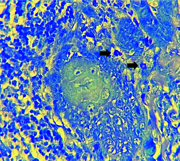
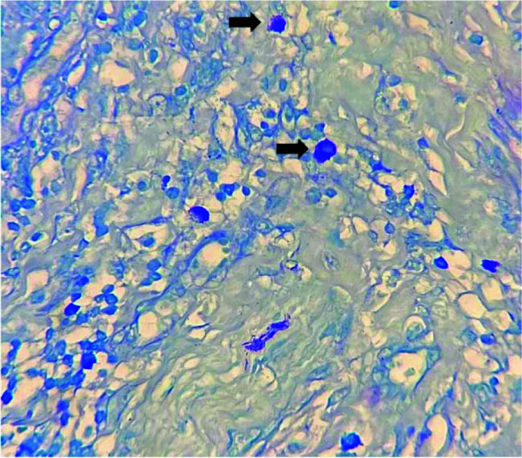
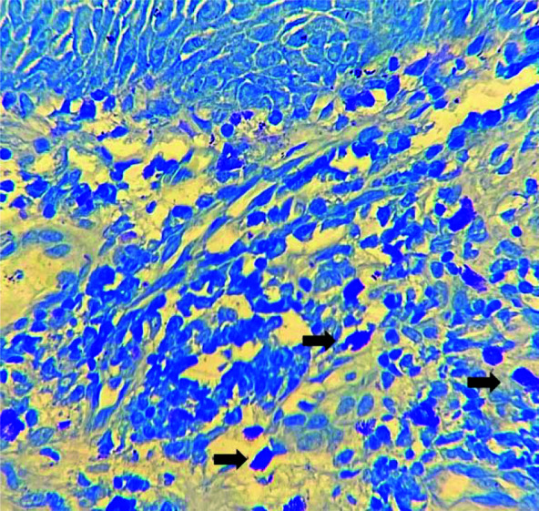
Materials and method: In this retrospective cross-sectional study, sixty-three archival cases, including 22 OSCCs, 28 dysplastic leukoplakias as epithelial dysplasia (ED), and 13 normal oral mucosal tissues, were examined for mast cells, using toluidine blue staining. Hotspot areas were identified at 10× magnification and mast cells were counted in 5 fields at 40× magnification. The average cell numbers were calculated, and the severity of inflammation was scored. Statistical analysis was performed using SPSS statistical software 20, including One-way ANOVA, Two-way ANOVA, paired-t test, and independent t-test. p Value < 0.05 was considered significant.
Results: Among the 51 pathologic lesions, 54.9% were males and 45.1% were females, with a mean age of 56.34±15.35 years. The most common locations were the tongue and buccal mucosa. The mast cell count was significantly lower in SCC compared to ED (p= 0.009). There was no correlation between mast cell count and inflammation score (p= 0.345).
Conclusion: In this study, the mast cell count was higher in ED compared to OSCC, suggesting an increase in these cells during the pre-malignant stages. However, the number of mast cells decreased after connective tissue invasion and microenvironmental changes occurred.


 分享
分享

 求助内容:
求助内容: 应助结果提醒方式:
 扫码关注我们
