{"title":"脂肪细胞对甲状旁腺瘤超声诊断的影响。","authors":"Tomoko Fujimoto, Mitsuyoshi Hirokawa, Ayana Suzuki, Maki Oshita, Hiroyuki Yamaoka, Makoto Fujishima, Naoyoshi Onoda, Akira Miyauchi, Takashi Akamizu","doi":"10.1007/s10396-024-01511-2","DOIUrl":null,"url":null,"abstract":"<p><strong>Purpose: </strong>Parathyroid lipoadenomas are difficult to recognize preoperatively; hence, they may remain undetected. Difficulty in recognition is thought to be due to the adipocytes present in the tumor. This study aimed to clarify the impact of adipocytes as a component of parathyroid adenomas on ultrasound evaluation.</p><p><strong>Methods: </strong>Eighteen parathyroid adenoma cases, in which the adipose tissue accounted for more than 10% of the tumors, were included in this study. Of these, five were consistent with lipoadenomas. Twenty-five consecutive patients with parathyroid adenoma without adipocytes were used as controls.</p><p><strong>Results: </strong>Ultrasonography revealed a lipoadenoma detection rate of 20.0%. This increased to 80.0% at re-examinations performed after obtaining information from other imaging modalities. Compared with parathyroid adenoma cases with no adipocytes or few adipocytes, the frequencies of ill-defined margins, iso- and/or hyperechogenicity, heterogeneous consistency with a two-tone pattern, poor vascular flow, no polar artery, and no hyperechoic line were significantly higher in parathyroid lipoadenoma cases. The hyperechoic and isoechoic areas in tumors with a two-tone pattern correspond to adipocyte- and parathyroid cell-rich areas, respectively. The lipoadenoma tumor sizes measured using ultrasound tended to be smaller than the actual sizes.</p><p><strong>Conclusions: </strong>The characteristic ultrasound findings of lipoadenomas were clearly different from those of parathyroid adenomas with or without adipocytes. We believe that our findings may contribute to an increased detection rate of lipoadenomas and allow us to consider them in the differential diagnosis.</p>","PeriodicalId":50130,"journal":{"name":"Journal of Medical Ultrasonics","volume":" ","pages":"237-243"},"PeriodicalIF":2.1000,"publicationDate":"2025-04-01","publicationTypes":"Journal Article","fieldsOfStudy":null,"isOpenAccess":false,"openAccessPdf":"https://www.ncbi.nlm.nih.gov/pmc/articles/PMC12018633/pdf/","citationCount":"0","resultStr":"{\"title\":\"Impact of adipocytes on ultrasound evaluation of parathyroid adenomas.\",\"authors\":\"Tomoko Fujimoto, Mitsuyoshi Hirokawa, Ayana Suzuki, Maki Oshita, Hiroyuki Yamaoka, Makoto Fujishima, Naoyoshi Onoda, Akira Miyauchi, Takashi Akamizu\",\"doi\":\"10.1007/s10396-024-01511-2\",\"DOIUrl\":null,\"url\":null,\"abstract\":\"<p><strong>Purpose: </strong>Parathyroid lipoadenomas are difficult to recognize preoperatively; hence, they may remain undetected. Difficulty in recognition is thought to be due to the adipocytes present in the tumor. This study aimed to clarify the impact of adipocytes as a component of parathyroid adenomas on ultrasound evaluation.</p><p><strong>Methods: </strong>Eighteen parathyroid adenoma cases, in which the adipose tissue accounted for more than 10% of the tumors, were included in this study. Of these, five were consistent with lipoadenomas. Twenty-five consecutive patients with parathyroid adenoma without adipocytes were used as controls.</p><p><strong>Results: </strong>Ultrasonography revealed a lipoadenoma detection rate of 20.0%. This increased to 80.0% at re-examinations performed after obtaining information from other imaging modalities. Compared with parathyroid adenoma cases with no adipocytes or few adipocytes, the frequencies of ill-defined margins, iso- and/or hyperechogenicity, heterogeneous consistency with a two-tone pattern, poor vascular flow, no polar artery, and no hyperechoic line were significantly higher in parathyroid lipoadenoma cases. The hyperechoic and isoechoic areas in tumors with a two-tone pattern correspond to adipocyte- and parathyroid cell-rich areas, respectively. The lipoadenoma tumor sizes measured using ultrasound tended to be smaller than the actual sizes.</p><p><strong>Conclusions: </strong>The characteristic ultrasound findings of lipoadenomas were clearly different from those of parathyroid adenomas with or without adipocytes. We believe that our findings may contribute to an increased detection rate of lipoadenomas and allow us to consider them in the differential diagnosis.</p>\",\"PeriodicalId\":50130,\"journal\":{\"name\":\"Journal of Medical Ultrasonics\",\"volume\":\" \",\"pages\":\"237-243\"},\"PeriodicalIF\":2.1000,\"publicationDate\":\"2025-04-01\",\"publicationTypes\":\"Journal Article\",\"fieldsOfStudy\":null,\"isOpenAccess\":false,\"openAccessPdf\":\"https://www.ncbi.nlm.nih.gov/pmc/articles/PMC12018633/pdf/\",\"citationCount\":\"0\",\"resultStr\":null,\"platform\":\"Semanticscholar\",\"paperid\":null,\"PeriodicalName\":\"Journal of Medical Ultrasonics\",\"FirstCategoryId\":\"3\",\"ListUrlMain\":\"https://doi.org/10.1007/s10396-024-01511-2\",\"RegionNum\":4,\"RegionCategory\":\"医学\",\"ArticlePicture\":[],\"TitleCN\":null,\"AbstractTextCN\":null,\"PMCID\":null,\"EPubDate\":\"2024/12/28 0:00:00\",\"PubModel\":\"Epub\",\"JCR\":\"Q3\",\"JCRName\":\"RADIOLOGY, NUCLEAR MEDICINE & MEDICAL IMAGING\",\"Score\":null,\"Total\":0}","platform":"Semanticscholar","paperid":null,"PeriodicalName":"Journal of Medical Ultrasonics","FirstCategoryId":"3","ListUrlMain":"https://doi.org/10.1007/s10396-024-01511-2","RegionNum":4,"RegionCategory":"医学","ArticlePicture":[],"TitleCN":null,"AbstractTextCN":null,"PMCID":null,"EPubDate":"2024/12/28 0:00:00","PubModel":"Epub","JCR":"Q3","JCRName":"RADIOLOGY, NUCLEAR MEDICINE & MEDICAL IMAGING","Score":null,"Total":0}
Impact of adipocytes on ultrasound evaluation of parathyroid adenomas.
Purpose: Parathyroid lipoadenomas are difficult to recognize preoperatively; hence, they may remain undetected. Difficulty in recognition is thought to be due to the adipocytes present in the tumor. This study aimed to clarify the impact of adipocytes as a component of parathyroid adenomas on ultrasound evaluation.
Methods: Eighteen parathyroid adenoma cases, in which the adipose tissue accounted for more than 10% of the tumors, were included in this study. Of these, five were consistent with lipoadenomas. Twenty-five consecutive patients with parathyroid adenoma without adipocytes were used as controls.
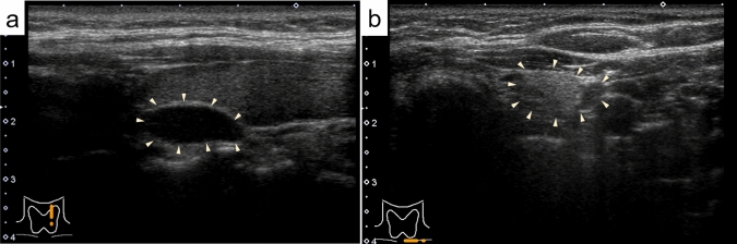
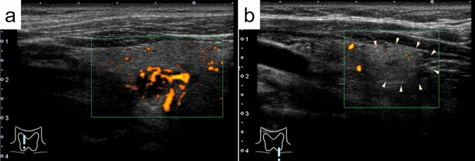
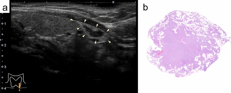
Results: Ultrasonography revealed a lipoadenoma detection rate of 20.0%. This increased to 80.0% at re-examinations performed after obtaining information from other imaging modalities. Compared with parathyroid adenoma cases with no adipocytes or few adipocytes, the frequencies of ill-defined margins, iso- and/or hyperechogenicity, heterogeneous consistency with a two-tone pattern, poor vascular flow, no polar artery, and no hyperechoic line were significantly higher in parathyroid lipoadenoma cases. The hyperechoic and isoechoic areas in tumors with a two-tone pattern correspond to adipocyte- and parathyroid cell-rich areas, respectively. The lipoadenoma tumor sizes measured using ultrasound tended to be smaller than the actual sizes.
Conclusions: The characteristic ultrasound findings of lipoadenomas were clearly different from those of parathyroid adenomas with or without adipocytes. We believe that our findings may contribute to an increased detection rate of lipoadenomas and allow us to consider them in the differential diagnosis.
期刊介绍:
The Journal of Medical Ultrasonics is the official journal of the Japan Society of Ultrasonics in Medicine. The main purpose of the journal is to provide forum for the publication of papers documenting recent advances and new developments in the entire field of ultrasound in medicine and biology, encompassing both the medical and the engineering aspects of the science.The journal welcomes original articles, review articles, images, and letters to the editor.The journal also provides state-of-the-art information such as announcements from the boards and the committees of the society.


 分享
分享
 求助内容:
求助内容: 应助结果提醒方式:
 扫码关注我们
