Mattin Sayed, Sari Saba-Sadiya, Benedikt Wichtlhuber, Julia Dietz, Matthias Neitzel, Leopold Keller, Gemma Roig, Andreas M Bucher
{"title":"利用增强技术评价医学图像分割模型。","authors":"Mattin Sayed, Sari Saba-Sadiya, Benedikt Wichtlhuber, Julia Dietz, Matthias Neitzel, Leopold Keller, Gemma Roig, Andreas M Bucher","doi":"10.3390/tomography10120150","DOIUrl":null,"url":null,"abstract":"<p><strong>Background: </strong>Medical imagesegmentation is an essential step in both clinical and research applications, and automated segmentation models-such as TotalSegmentator-have become ubiquitous. However, robust methods for validating the accuracy of these models remain limited, and manual inspection is often necessary before the segmentation masks produced by these models can be used.</p><p><strong>Methods: </strong>To address this gap, we have developed a novel validation framework for segmentation models, leveraging data augmentation to assess model consistency. We produced segmentation masks for both the original and augmented scans, and we calculated the alignment metrics between these segmentation masks.</p><p><strong>Results: </strong>Our results demonstrate strong correlation between the segmentation quality of the original scan and the average alignment between the masks of the original and augmented CT scans. These results were further validated by supporting metrics, including the coefficient of variance and the average symmetric surface distance, indicating that agreement with augmented-scan segmentation masks is a valid proxy for segmentation quality.</p><p><strong>Conclusions: </strong>Overall, our framework offers a pipeline for evaluating segmentation performance without relying on manually labeled ground truth data, establishing a foundation for future advancements in automated medical image analysis.</p>","PeriodicalId":51330,"journal":{"name":"Tomography","volume":"10 12","pages":"2128-2143"},"PeriodicalIF":2.2000,"publicationDate":"2024-12-23","publicationTypes":"Journal Article","fieldsOfStudy":null,"isOpenAccess":false,"openAccessPdf":"https://www.ncbi.nlm.nih.gov/pmc/articles/PMC11679113/pdf/","citationCount":"0","resultStr":"{\"title\":\"Evaluating Medical Image Segmentation Models Using Augmentation.\",\"authors\":\"Mattin Sayed, Sari Saba-Sadiya, Benedikt Wichtlhuber, Julia Dietz, Matthias Neitzel, Leopold Keller, Gemma Roig, Andreas M Bucher\",\"doi\":\"10.3390/tomography10120150\",\"DOIUrl\":null,\"url\":null,\"abstract\":\"<p><strong>Background: </strong>Medical imagesegmentation is an essential step in both clinical and research applications, and automated segmentation models-such as TotalSegmentator-have become ubiquitous. However, robust methods for validating the accuracy of these models remain limited, and manual inspection is often necessary before the segmentation masks produced by these models can be used.</p><p><strong>Methods: </strong>To address this gap, we have developed a novel validation framework for segmentation models, leveraging data augmentation to assess model consistency. We produced segmentation masks for both the original and augmented scans, and we calculated the alignment metrics between these segmentation masks.</p><p><strong>Results: </strong>Our results demonstrate strong correlation between the segmentation quality of the original scan and the average alignment between the masks of the original and augmented CT scans. These results were further validated by supporting metrics, including the coefficient of variance and the average symmetric surface distance, indicating that agreement with augmented-scan segmentation masks is a valid proxy for segmentation quality.</p><p><strong>Conclusions: </strong>Overall, our framework offers a pipeline for evaluating segmentation performance without relying on manually labeled ground truth data, establishing a foundation for future advancements in automated medical image analysis.</p>\",\"PeriodicalId\":51330,\"journal\":{\"name\":\"Tomography\",\"volume\":\"10 12\",\"pages\":\"2128-2143\"},\"PeriodicalIF\":2.2000,\"publicationDate\":\"2024-12-23\",\"publicationTypes\":\"Journal Article\",\"fieldsOfStudy\":null,\"isOpenAccess\":false,\"openAccessPdf\":\"https://www.ncbi.nlm.nih.gov/pmc/articles/PMC11679113/pdf/\",\"citationCount\":\"0\",\"resultStr\":null,\"platform\":\"Semanticscholar\",\"paperid\":null,\"PeriodicalName\":\"Tomography\",\"FirstCategoryId\":\"3\",\"ListUrlMain\":\"https://doi.org/10.3390/tomography10120150\",\"RegionNum\":4,\"RegionCategory\":\"医学\",\"ArticlePicture\":[],\"TitleCN\":null,\"AbstractTextCN\":null,\"PMCID\":null,\"EPubDate\":\"\",\"PubModel\":\"\",\"JCR\":\"Q2\",\"JCRName\":\"RADIOLOGY, NUCLEAR MEDICINE & MEDICAL IMAGING\",\"Score\":null,\"Total\":0}","platform":"Semanticscholar","paperid":null,"PeriodicalName":"Tomography","FirstCategoryId":"3","ListUrlMain":"https://doi.org/10.3390/tomography10120150","RegionNum":4,"RegionCategory":"医学","ArticlePicture":[],"TitleCN":null,"AbstractTextCN":null,"PMCID":null,"EPubDate":"","PubModel":"","JCR":"Q2","JCRName":"RADIOLOGY, NUCLEAR MEDICINE & MEDICAL IMAGING","Score":null,"Total":0}
Evaluating Medical Image Segmentation Models Using Augmentation.
Background: Medical imagesegmentation is an essential step in both clinical and research applications, and automated segmentation models-such as TotalSegmentator-have become ubiquitous. However, robust methods for validating the accuracy of these models remain limited, and manual inspection is often necessary before the segmentation masks produced by these models can be used.
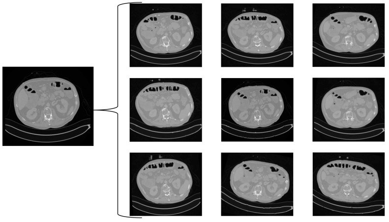
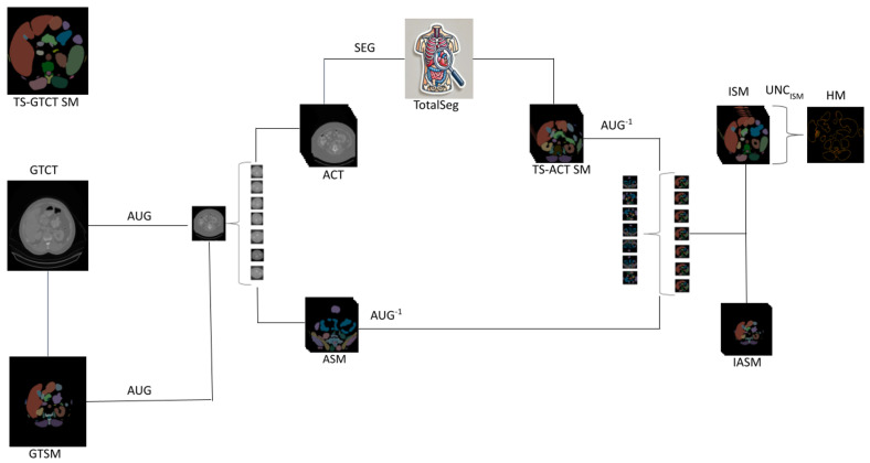
Methods: To address this gap, we have developed a novel validation framework for segmentation models, leveraging data augmentation to assess model consistency. We produced segmentation masks for both the original and augmented scans, and we calculated the alignment metrics between these segmentation masks.
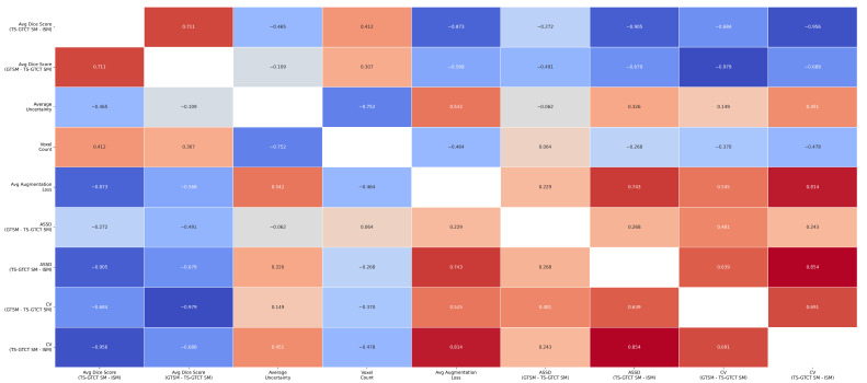
Results: Our results demonstrate strong correlation between the segmentation quality of the original scan and the average alignment between the masks of the original and augmented CT scans. These results were further validated by supporting metrics, including the coefficient of variance and the average symmetric surface distance, indicating that agreement with augmented-scan segmentation masks is a valid proxy for segmentation quality.
Conclusions: Overall, our framework offers a pipeline for evaluating segmentation performance without relying on manually labeled ground truth data, establishing a foundation for future advancements in automated medical image analysis.
TomographyMedicine-Radiology, Nuclear Medicine and Imaging
CiteScore
2.70
自引率
10.50%
发文量
222
期刊介绍:
TomographyTM publishes basic (technical and pre-clinical) and clinical scientific articles which involve the advancement of imaging technologies. Tomography encompasses studies that use single or multiple imaging modalities including for example CT, US, PET, SPECT, MR and hyperpolarization technologies, as well as optical modalities (i.e. bioluminescence, photoacoustic, endomicroscopy, fiber optic imaging and optical computed tomography) in basic sciences, engineering, preclinical and clinical medicine.
Tomography also welcomes studies involving exploration and refinement of contrast mechanisms and image-derived metrics within and across modalities toward the development of novel imaging probes for image-based feedback and intervention. The use of imaging in biology and medicine provides unparalleled opportunities to noninvasively interrogate tissues to obtain real-time dynamic and quantitative information required for diagnosis and response to interventions and to follow evolving pathological conditions. As multi-modal studies and the complexities of imaging technologies themselves are ever increasing to provide advanced information to scientists and clinicians.
Tomography provides a unique publication venue allowing investigators the opportunity to more precisely communicate integrated findings related to the diverse and heterogeneous features associated with underlying anatomical, physiological, functional, metabolic and molecular genetic activities of normal and diseased tissue. Thus Tomography publishes peer-reviewed articles which involve the broad use of imaging of any tissue and disease type including both preclinical and clinical investigations. In addition, hardware/software along with chemical and molecular probe advances are welcome as they are deemed to significantly contribute towards the long-term goal of improving the overall impact of imaging on scientific and clinical discovery.


 分享
分享
 求助内容:
求助内容: 应助结果提醒方式:
 扫码关注我们
