Takashi Okazaki, Tetsu Niwa, Ryoichi Yoshida, Takatoshi Sorimachi, Jun Hashimoto
{"title":"使用超高分辨率光子计数检测器计算机断层扫描(CT)血管造影术观察颅内穿动脉。","authors":"Takashi Okazaki, Tetsu Niwa, Ryoichi Yoshida, Takatoshi Sorimachi, Jun Hashimoto","doi":"10.3390/tomography10120136","DOIUrl":null,"url":null,"abstract":"<p><p><b>Background/Objectives:</b> Photon-counting detector computed tomography (PCD-CT) offers energy-resolved CT data with enhanced resolution, reduced electronic noise, and improved tissue contrast. This study aimed to evaluate the visibility of intracranial perforating arteries on ultra-high-resolution (UHR) CT angiography (CTA) on PCD-CT. <b>Methods:</b> A retrospective analysis of intracranial UHR PCD-CTA was performed for 30 patients. The image quality from four UHR PCD-CTA reconstruction methods [kernel Hv40 and Hv72, with and without quantum iterative reconstruction (QIR)] was assessed for the lenticulostriate arteries (LSAs) and pontine arteries (PAs). A subjective evaluation included peripheral visibility, vessel sharpness, and image noise, while objective analysis focused on the signal-to-noise ratio (SNR) and contrast-to-noise ratio (CNR). <b>Results:</b> Peripheral LSAs were well visualized across all reconstruction methods, with no significant differences between them. Vessel sharpness and image noise varied significantly (<i>p</i> < 0.0001); sharper LSAs and more noise were seen with kernel Hv72 compared to kernel Hv40 (<i>p</i> < 0.05). A similar pattern was observed for PAs, though peripheral visibility was lower than that for LSAs. The SNR and CNR were the highest in the presence of kernel Hv72 with QIR, and lowest with kernel Hv72 without QIR, compared to kernel Hv40 (<i>p</i> < 0.05). <b>Conclusions:</b> UHR PCD-CTA provided a good visualization of the intracranial perforating arteries, particularly LSAs. The vessel sharpness and image noise varied by reconstruction method, in which kernel Hv72 with QIR offered the optimal visualization.</p>","PeriodicalId":51330,"journal":{"name":"Tomography","volume":"10 12","pages":"1867-1880"},"PeriodicalIF":2.2000,"publicationDate":"2024-11-21","publicationTypes":"Journal Article","fieldsOfStudy":null,"isOpenAccess":false,"openAccessPdf":"https://www.ncbi.nlm.nih.gov/pmc/articles/PMC11679214/pdf/","citationCount":"0","resultStr":"{\"title\":\"Visibility of Intracranial Perforating Arteries Using Ultra-High-Resolution Photon-Counting Detector Computed Tomography (CT) Angiography.\",\"authors\":\"Takashi Okazaki, Tetsu Niwa, Ryoichi Yoshida, Takatoshi Sorimachi, Jun Hashimoto\",\"doi\":\"10.3390/tomography10120136\",\"DOIUrl\":null,\"url\":null,\"abstract\":\"<p><p><b>Background/Objectives:</b> Photon-counting detector computed tomography (PCD-CT) offers energy-resolved CT data with enhanced resolution, reduced electronic noise, and improved tissue contrast. This study aimed to evaluate the visibility of intracranial perforating arteries on ultra-high-resolution (UHR) CT angiography (CTA) on PCD-CT. <b>Methods:</b> A retrospective analysis of intracranial UHR PCD-CTA was performed for 30 patients. The image quality from four UHR PCD-CTA reconstruction methods [kernel Hv40 and Hv72, with and without quantum iterative reconstruction (QIR)] was assessed for the lenticulostriate arteries (LSAs) and pontine arteries (PAs). A subjective evaluation included peripheral visibility, vessel sharpness, and image noise, while objective analysis focused on the signal-to-noise ratio (SNR) and contrast-to-noise ratio (CNR). <b>Results:</b> Peripheral LSAs were well visualized across all reconstruction methods, with no significant differences between them. Vessel sharpness and image noise varied significantly (<i>p</i> < 0.0001); sharper LSAs and more noise were seen with kernel Hv72 compared to kernel Hv40 (<i>p</i> < 0.05). A similar pattern was observed for PAs, though peripheral visibility was lower than that for LSAs. The SNR and CNR were the highest in the presence of kernel Hv72 with QIR, and lowest with kernel Hv72 without QIR, compared to kernel Hv40 (<i>p</i> < 0.05). <b>Conclusions:</b> UHR PCD-CTA provided a good visualization of the intracranial perforating arteries, particularly LSAs. The vessel sharpness and image noise varied by reconstruction method, in which kernel Hv72 with QIR offered the optimal visualization.</p>\",\"PeriodicalId\":51330,\"journal\":{\"name\":\"Tomography\",\"volume\":\"10 12\",\"pages\":\"1867-1880\"},\"PeriodicalIF\":2.2000,\"publicationDate\":\"2024-11-21\",\"publicationTypes\":\"Journal Article\",\"fieldsOfStudy\":null,\"isOpenAccess\":false,\"openAccessPdf\":\"https://www.ncbi.nlm.nih.gov/pmc/articles/PMC11679214/pdf/\",\"citationCount\":\"0\",\"resultStr\":null,\"platform\":\"Semanticscholar\",\"paperid\":null,\"PeriodicalName\":\"Tomography\",\"FirstCategoryId\":\"3\",\"ListUrlMain\":\"https://doi.org/10.3390/tomography10120136\",\"RegionNum\":4,\"RegionCategory\":\"医学\",\"ArticlePicture\":[],\"TitleCN\":null,\"AbstractTextCN\":null,\"PMCID\":null,\"EPubDate\":\"\",\"PubModel\":\"\",\"JCR\":\"Q2\",\"JCRName\":\"RADIOLOGY, NUCLEAR MEDICINE & MEDICAL IMAGING\",\"Score\":null,\"Total\":0}","platform":"Semanticscholar","paperid":null,"PeriodicalName":"Tomography","FirstCategoryId":"3","ListUrlMain":"https://doi.org/10.3390/tomography10120136","RegionNum":4,"RegionCategory":"医学","ArticlePicture":[],"TitleCN":null,"AbstractTextCN":null,"PMCID":null,"EPubDate":"","PubModel":"","JCR":"Q2","JCRName":"RADIOLOGY, NUCLEAR MEDICINE & MEDICAL IMAGING","Score":null,"Total":0}
Visibility of Intracranial Perforating Arteries Using Ultra-High-Resolution Photon-Counting Detector Computed Tomography (CT) Angiography.
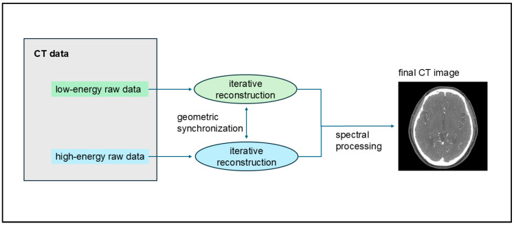
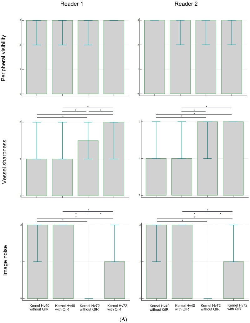
Background/Objectives: Photon-counting detector computed tomography (PCD-CT) offers energy-resolved CT data with enhanced resolution, reduced electronic noise, and improved tissue contrast. This study aimed to evaluate the visibility of intracranial perforating arteries on ultra-high-resolution (UHR) CT angiography (CTA) on PCD-CT. Methods: A retrospective analysis of intracranial UHR PCD-CTA was performed for 30 patients. The image quality from four UHR PCD-CTA reconstruction methods [kernel Hv40 and Hv72, with and without quantum iterative reconstruction (QIR)] was assessed for the lenticulostriate arteries (LSAs) and pontine arteries (PAs). A subjective evaluation included peripheral visibility, vessel sharpness, and image noise, while objective analysis focused on the signal-to-noise ratio (SNR) and contrast-to-noise ratio (CNR). Results: Peripheral LSAs were well visualized across all reconstruction methods, with no significant differences between them. Vessel sharpness and image noise varied significantly (p < 0.0001); sharper LSAs and more noise were seen with kernel Hv72 compared to kernel Hv40 (p < 0.05). A similar pattern was observed for PAs, though peripheral visibility was lower than that for LSAs. The SNR and CNR were the highest in the presence of kernel Hv72 with QIR, and lowest with kernel Hv72 without QIR, compared to kernel Hv40 (p < 0.05). Conclusions: UHR PCD-CTA provided a good visualization of the intracranial perforating arteries, particularly LSAs. The vessel sharpness and image noise varied by reconstruction method, in which kernel Hv72 with QIR offered the optimal visualization.
TomographyMedicine-Radiology, Nuclear Medicine and Imaging
CiteScore
2.70
自引率
10.50%
发文量
222
期刊介绍:
TomographyTM publishes basic (technical and pre-clinical) and clinical scientific articles which involve the advancement of imaging technologies. Tomography encompasses studies that use single or multiple imaging modalities including for example CT, US, PET, SPECT, MR and hyperpolarization technologies, as well as optical modalities (i.e. bioluminescence, photoacoustic, endomicroscopy, fiber optic imaging and optical computed tomography) in basic sciences, engineering, preclinical and clinical medicine.
Tomography also welcomes studies involving exploration and refinement of contrast mechanisms and image-derived metrics within and across modalities toward the development of novel imaging probes for image-based feedback and intervention. The use of imaging in biology and medicine provides unparalleled opportunities to noninvasively interrogate tissues to obtain real-time dynamic and quantitative information required for diagnosis and response to interventions and to follow evolving pathological conditions. As multi-modal studies and the complexities of imaging technologies themselves are ever increasing to provide advanced information to scientists and clinicians.
Tomography provides a unique publication venue allowing investigators the opportunity to more precisely communicate integrated findings related to the diverse and heterogeneous features associated with underlying anatomical, physiological, functional, metabolic and molecular genetic activities of normal and diseased tissue. Thus Tomography publishes peer-reviewed articles which involve the broad use of imaging of any tissue and disease type including both preclinical and clinical investigations. In addition, hardware/software along with chemical and molecular probe advances are welcome as they are deemed to significantly contribute towards the long-term goal of improving the overall impact of imaging on scientific and clinical discovery.


 分享
分享
 求助内容:
求助内容: 应助结果提醒方式:
 扫码关注我们
