Oguz Karcioglu, Ayşen Kara, Olcay Kurtulan, Serkan Uysal, Ziya Toros Selçuk
{"title":"支气管镜下肺包虫囊提取术及手术必要性1例。","authors":"Oguz Karcioglu, Ayşen Kara, Olcay Kurtulan, Serkan Uysal, Ziya Toros Selçuk","doi":"10.18502/ijpa.v19i4.17170","DOIUrl":null,"url":null,"abstract":"<p><p><i>Echinococcus granulosus</i> remains a global public health issue. Although predominantly affecting the liver, the lungs are the second most affected organ and often undergo surgical intervention. Here, a case managed by bronchoscopy and medical therapy is presented. A 26-year-old woman was presented with a cough, hemoptysis, and a 5 kg weight loss in the last two months. Chest imaging identified a 4 cm centrally cystic mass lesion in the middle lobe of the right lung, which was suspicious of lung cancer. Bronchoscopy revealed a whitish, plastic-like object that was difficult to extricate and obstructed the middle lobe bronchus. We removed the material and purulent secretions covering it and opened the middle lobe bronchus totally. The histopathological study verified its consistency with hydatid cyst. There was no evidence of a hydatid cyst on computerized thomography after bronchoscopy. The lesion in the left lobe of the liver, confirmed to be suggestive of a hydatid cyst via ultrasonography, was treated using the PAIR technique. We administered oral albendazole to continue the treatment. It may be a reasonable approach to postpone surgery in order to preserve lung tissue in patients who have undergone complete removal of hydatid cyst material via bronchoscope.</p>","PeriodicalId":14669,"journal":{"name":"Iranian Journal of Parasitology","volume":"19 4","pages":"484-488"},"PeriodicalIF":0.9000,"publicationDate":"2024-10-01","publicationTypes":"Journal Article","fieldsOfStudy":null,"isOpenAccess":false,"openAccessPdf":"https://www.ncbi.nlm.nih.gov/pmc/articles/PMC11671833/pdf/","citationCount":"0","resultStr":"{\"title\":\"Lung Cyst Hydatid Extracted via Bronchoscopy and the Necessity of Surgery: A Case Report.\",\"authors\":\"Oguz Karcioglu, Ayşen Kara, Olcay Kurtulan, Serkan Uysal, Ziya Toros Selçuk\",\"doi\":\"10.18502/ijpa.v19i4.17170\",\"DOIUrl\":null,\"url\":null,\"abstract\":\"<p><p><i>Echinococcus granulosus</i> remains a global public health issue. Although predominantly affecting the liver, the lungs are the second most affected organ and often undergo surgical intervention. Here, a case managed by bronchoscopy and medical therapy is presented. A 26-year-old woman was presented with a cough, hemoptysis, and a 5 kg weight loss in the last two months. Chest imaging identified a 4 cm centrally cystic mass lesion in the middle lobe of the right lung, which was suspicious of lung cancer. Bronchoscopy revealed a whitish, plastic-like object that was difficult to extricate and obstructed the middle lobe bronchus. We removed the material and purulent secretions covering it and opened the middle lobe bronchus totally. The histopathological study verified its consistency with hydatid cyst. There was no evidence of a hydatid cyst on computerized thomography after bronchoscopy. The lesion in the left lobe of the liver, confirmed to be suggestive of a hydatid cyst via ultrasonography, was treated using the PAIR technique. We administered oral albendazole to continue the treatment. It may be a reasonable approach to postpone surgery in order to preserve lung tissue in patients who have undergone complete removal of hydatid cyst material via bronchoscope.</p>\",\"PeriodicalId\":14669,\"journal\":{\"name\":\"Iranian Journal of Parasitology\",\"volume\":\"19 4\",\"pages\":\"484-488\"},\"PeriodicalIF\":0.9000,\"publicationDate\":\"2024-10-01\",\"publicationTypes\":\"Journal Article\",\"fieldsOfStudy\":null,\"isOpenAccess\":false,\"openAccessPdf\":\"https://www.ncbi.nlm.nih.gov/pmc/articles/PMC11671833/pdf/\",\"citationCount\":\"0\",\"resultStr\":null,\"platform\":\"Semanticscholar\",\"paperid\":null,\"PeriodicalName\":\"Iranian Journal of Parasitology\",\"FirstCategoryId\":\"3\",\"ListUrlMain\":\"https://doi.org/10.18502/ijpa.v19i4.17170\",\"RegionNum\":4,\"RegionCategory\":\"医学\",\"ArticlePicture\":[],\"TitleCN\":null,\"AbstractTextCN\":null,\"PMCID\":null,\"EPubDate\":\"\",\"PubModel\":\"\",\"JCR\":\"Q4\",\"JCRName\":\"PARASITOLOGY\",\"Score\":null,\"Total\":0}","platform":"Semanticscholar","paperid":null,"PeriodicalName":"Iranian Journal of Parasitology","FirstCategoryId":"3","ListUrlMain":"https://doi.org/10.18502/ijpa.v19i4.17170","RegionNum":4,"RegionCategory":"医学","ArticlePicture":[],"TitleCN":null,"AbstractTextCN":null,"PMCID":null,"EPubDate":"","PubModel":"","JCR":"Q4","JCRName":"PARASITOLOGY","Score":null,"Total":0}
Lung Cyst Hydatid Extracted via Bronchoscopy and the Necessity of Surgery: A Case Report.
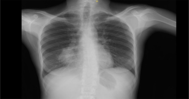
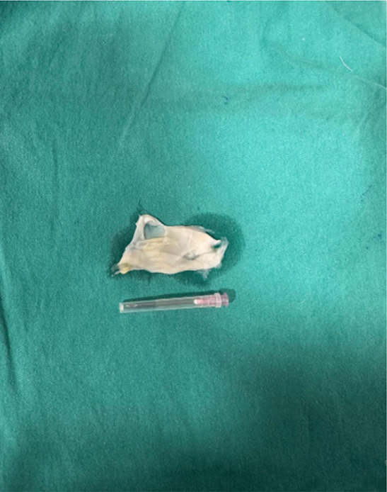
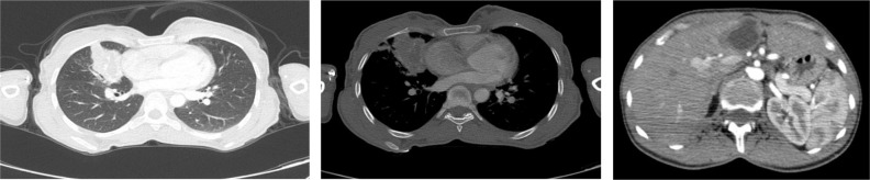
Echinococcus granulosus remains a global public health issue. Although predominantly affecting the liver, the lungs are the second most affected organ and often undergo surgical intervention. Here, a case managed by bronchoscopy and medical therapy is presented. A 26-year-old woman was presented with a cough, hemoptysis, and a 5 kg weight loss in the last two months. Chest imaging identified a 4 cm centrally cystic mass lesion in the middle lobe of the right lung, which was suspicious of lung cancer. Bronchoscopy revealed a whitish, plastic-like object that was difficult to extricate and obstructed the middle lobe bronchus. We removed the material and purulent secretions covering it and opened the middle lobe bronchus totally. The histopathological study verified its consistency with hydatid cyst. There was no evidence of a hydatid cyst on computerized thomography after bronchoscopy. The lesion in the left lobe of the liver, confirmed to be suggestive of a hydatid cyst via ultrasonography, was treated using the PAIR technique. We administered oral albendazole to continue the treatment. It may be a reasonable approach to postpone surgery in order to preserve lung tissue in patients who have undergone complete removal of hydatid cyst material via bronchoscope.
期刊介绍:
Iranian Journal of Parasitology (IJP) is the official publication of Iranian Society of Parasitology (ISP) launched in 2006. The society was inaugurated in 1994 and pursues the improvement of the knowledge on the parasites and parasitic diseases, exchange of scientific knowledge with foreign societies, publicity activities, and consultation on the parasitic diseases, and intimate relationship among society members.
The main aims of the Journal are: contribution to the field of Parasitology, including all aspects of parasites and parasitic diseases (medical and veterinary) and related fields such as Entomology which may be submitted by scientists from Iran and all over the world.


 分享
分享
 求助内容:
求助内容: 应助结果提醒方式:
 扫码关注我们
