{"title":"急诊科一例罕见的低视力和斜视的神经学发现:眼色素沉着症。","authors":"Osman Özen, Menekşe İnal Özen","doi":"10.4274/tjo.galenos.2024.77783","DOIUrl":null,"url":null,"abstract":"<p><p>We present the case of a patient who came to the emergency department with a significant decrease in vision and dilated pupil in the left eye. Since neurological pathologies were primarily considered, diffusion brain magnetic resonance imaging (MRI) and brain computed tomography (CT) were requested. After the results were reported as normal, we were consulted. On examination, the anterior segment was normal but we detected shiny pearl-like formations in the anterior vitreous, condensation at the inferior of the posterior vitreous, and a scar in the macula. When we evaluated the orbital section of the current brain CT, we detected an intraocular foreign body (IOFB). On the brain MRI, we saw a large artifact that obscured the left orbit and surrounding anatomical structures. When we questioned again, we learned that he had been admitted to another emergency department two months prior due to an object hitting his left eye, where the eye was only washed with saline. Our case emphasizes that ocular siderosis caused by IOFBs should be kept in mind in the differential diagnosis of anisocoria, especially before MRI. Because metallic objects may move during MRI, undiagnosed IOFBs can cause serious ocular side effects.</p>","PeriodicalId":23373,"journal":{"name":"Turkish Journal of Ophthalmology","volume":"54 6","pages":"354-357"},"PeriodicalIF":0.0000,"publicationDate":"2024-12-31","publicationTypes":"Journal Article","fieldsOfStudy":null,"isOpenAccess":false,"openAccessPdf":"https://www.ncbi.nlm.nih.gov/pmc/articles/PMC11707453/pdf/","citationCount":"0","resultStr":"{\"title\":\"An Unusual Case of Low Vision and Anisocoria Considered a Neurological Finding in the Emergency Department: Ocular Siderosis.\",\"authors\":\"Osman Özen, Menekşe İnal Özen\",\"doi\":\"10.4274/tjo.galenos.2024.77783\",\"DOIUrl\":null,\"url\":null,\"abstract\":\"<p><p>We present the case of a patient who came to the emergency department with a significant decrease in vision and dilated pupil in the left eye. Since neurological pathologies were primarily considered, diffusion brain magnetic resonance imaging (MRI) and brain computed tomography (CT) were requested. After the results were reported as normal, we were consulted. On examination, the anterior segment was normal but we detected shiny pearl-like formations in the anterior vitreous, condensation at the inferior of the posterior vitreous, and a scar in the macula. When we evaluated the orbital section of the current brain CT, we detected an intraocular foreign body (IOFB). On the brain MRI, we saw a large artifact that obscured the left orbit and surrounding anatomical structures. When we questioned again, we learned that he had been admitted to another emergency department two months prior due to an object hitting his left eye, where the eye was only washed with saline. Our case emphasizes that ocular siderosis caused by IOFBs should be kept in mind in the differential diagnosis of anisocoria, especially before MRI. Because metallic objects may move during MRI, undiagnosed IOFBs can cause serious ocular side effects.</p>\",\"PeriodicalId\":23373,\"journal\":{\"name\":\"Turkish Journal of Ophthalmology\",\"volume\":\"54 6\",\"pages\":\"354-357\"},\"PeriodicalIF\":0.0000,\"publicationDate\":\"2024-12-31\",\"publicationTypes\":\"Journal Article\",\"fieldsOfStudy\":null,\"isOpenAccess\":false,\"openAccessPdf\":\"https://www.ncbi.nlm.nih.gov/pmc/articles/PMC11707453/pdf/\",\"citationCount\":\"0\",\"resultStr\":null,\"platform\":\"Semanticscholar\",\"paperid\":null,\"PeriodicalName\":\"Turkish Journal of Ophthalmology\",\"FirstCategoryId\":\"1085\",\"ListUrlMain\":\"https://doi.org/10.4274/tjo.galenos.2024.77783\",\"RegionNum\":0,\"RegionCategory\":null,\"ArticlePicture\":[],\"TitleCN\":null,\"AbstractTextCN\":null,\"PMCID\":null,\"EPubDate\":\"\",\"PubModel\":\"\",\"JCR\":\"Q3\",\"JCRName\":\"Medicine\",\"Score\":null,\"Total\":0}","platform":"Semanticscholar","paperid":null,"PeriodicalName":"Turkish Journal of Ophthalmology","FirstCategoryId":"1085","ListUrlMain":"https://doi.org/10.4274/tjo.galenos.2024.77783","RegionNum":0,"RegionCategory":null,"ArticlePicture":[],"TitleCN":null,"AbstractTextCN":null,"PMCID":null,"EPubDate":"","PubModel":"","JCR":"Q3","JCRName":"Medicine","Score":null,"Total":0}
An Unusual Case of Low Vision and Anisocoria Considered a Neurological Finding in the Emergency Department: Ocular Siderosis.
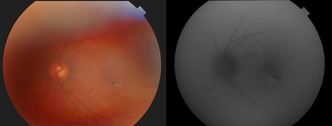
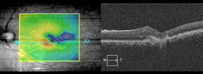
We present the case of a patient who came to the emergency department with a significant decrease in vision and dilated pupil in the left eye. Since neurological pathologies were primarily considered, diffusion brain magnetic resonance imaging (MRI) and brain computed tomography (CT) were requested. After the results were reported as normal, we were consulted. On examination, the anterior segment was normal but we detected shiny pearl-like formations in the anterior vitreous, condensation at the inferior of the posterior vitreous, and a scar in the macula. When we evaluated the orbital section of the current brain CT, we detected an intraocular foreign body (IOFB). On the brain MRI, we saw a large artifact that obscured the left orbit and surrounding anatomical structures. When we questioned again, we learned that he had been admitted to another emergency department two months prior due to an object hitting his left eye, where the eye was only washed with saline. Our case emphasizes that ocular siderosis caused by IOFBs should be kept in mind in the differential diagnosis of anisocoria, especially before MRI. Because metallic objects may move during MRI, undiagnosed IOFBs can cause serious ocular side effects.
期刊介绍:
The Turkish Journal of Ophthalmology (TJO) is the only scientific periodical publication of the Turkish Ophthalmological Association and has been published since January 1929. In its early years, the journal was published in Turkish and French. Although there were temporary interruptions in the publication of the journal due to various challenges, the Turkish Journal of Ophthalmology has been published continually from 1971 to the present. The target audience includes specialists and physicians in training in ophthalmology in all relevant disciplines.


 分享
分享
 求助内容:
求助内容: 应助结果提醒方式:
 扫码关注我们
