{"title":"腕管综合征和对照组正中神经磁共振成像的评价。","authors":"Ghasem Farahmand, Atefeh Behkar, Hassan Hashemi, Mahsa Ghajarzadeh, Samira Raminfard, Mojtaba Shahbazi, Payam Sarraf","doi":"10.18502/cjn.v23i2.16837","DOIUrl":null,"url":null,"abstract":"<p><p><b>Background:</b> Carpal tunnel syndrome (CTS) is a common peripheral nerve entrapment disorder that is diagnosed using clinical signs and symptoms and confirmed via nerve conduction studies (NCSs). While NCS is a semi-invasive procedure, magnetic resonance imaging (MRI) is a non-invasive diagnostic tool that detects macroscopic nerve abnormalities and evaluates a patient's surgical or medication treatment options. This study assessed magnetic resonance neurography (MRN)'s diagnostic and grading value by comparing it to electrodiagnostic studies in patients with CTS and healthy individuals. <b>Methods:</b> This was a cross-sectional study on 27 wrists with CTS and 27 healthy wrists. After history taking and physical examination, we employed an NCS to confirm and determine the severity of CTS, then MRN and diffusion tensor imaging (DTI) were used to calculate apparent diffusion coefficient (ADC), fractional anisotropy (FA), and cross-sectional area (CSA). <b>Results:</b> 18 patients with CTS (27 median nerves) and 15 healthy controls (27 median nerves) were evaluated. The mean FA in the CTS group was significantly lower (0.38 ± 0.05 vs. 0.45 ± 0.06, P < 0.001). The mean CSA and ADC were higher in patients with CTS but not statistically significant. FA's diagnostic cut-off was 0.42, with a sensitivity of 70.4% and a specificity of 63%. <b>Conclusion:</b> MRN with DTI can be an effective and non-invasive diagnostic technique for the detection of CTS. The FA measure demonstrated adequate sensitivity and specificity for differentiating patients with CTS from healthy individuals.</p>","PeriodicalId":40077,"journal":{"name":"Current Journal of Neurology","volume":"23 2","pages":"89-95"},"PeriodicalIF":0.5000,"publicationDate":"2024-04-03","publicationTypes":"Journal Article","fieldsOfStudy":null,"isOpenAccess":false,"openAccessPdf":"https://www.ncbi.nlm.nih.gov/pmc/articles/PMC11685557/pdf/","citationCount":"0","resultStr":"{\"title\":\"Assessment of median nerve with magnetic resonance neurography in cases with carpal tunnel syndrome and controls.\",\"authors\":\"Ghasem Farahmand, Atefeh Behkar, Hassan Hashemi, Mahsa Ghajarzadeh, Samira Raminfard, Mojtaba Shahbazi, Payam Sarraf\",\"doi\":\"10.18502/cjn.v23i2.16837\",\"DOIUrl\":null,\"url\":null,\"abstract\":\"<p><p><b>Background:</b> Carpal tunnel syndrome (CTS) is a common peripheral nerve entrapment disorder that is diagnosed using clinical signs and symptoms and confirmed via nerve conduction studies (NCSs). While NCS is a semi-invasive procedure, magnetic resonance imaging (MRI) is a non-invasive diagnostic tool that detects macroscopic nerve abnormalities and evaluates a patient's surgical or medication treatment options. This study assessed magnetic resonance neurography (MRN)'s diagnostic and grading value by comparing it to electrodiagnostic studies in patients with CTS and healthy individuals. <b>Methods:</b> This was a cross-sectional study on 27 wrists with CTS and 27 healthy wrists. After history taking and physical examination, we employed an NCS to confirm and determine the severity of CTS, then MRN and diffusion tensor imaging (DTI) were used to calculate apparent diffusion coefficient (ADC), fractional anisotropy (FA), and cross-sectional area (CSA). <b>Results:</b> 18 patients with CTS (27 median nerves) and 15 healthy controls (27 median nerves) were evaluated. The mean FA in the CTS group was significantly lower (0.38 ± 0.05 vs. 0.45 ± 0.06, P < 0.001). The mean CSA and ADC were higher in patients with CTS but not statistically significant. FA's diagnostic cut-off was 0.42, with a sensitivity of 70.4% and a specificity of 63%. <b>Conclusion:</b> MRN with DTI can be an effective and non-invasive diagnostic technique for the detection of CTS. The FA measure demonstrated adequate sensitivity and specificity for differentiating patients with CTS from healthy individuals.</p>\",\"PeriodicalId\":40077,\"journal\":{\"name\":\"Current Journal of Neurology\",\"volume\":\"23 2\",\"pages\":\"89-95\"},\"PeriodicalIF\":0.5000,\"publicationDate\":\"2024-04-03\",\"publicationTypes\":\"Journal Article\",\"fieldsOfStudy\":null,\"isOpenAccess\":false,\"openAccessPdf\":\"https://www.ncbi.nlm.nih.gov/pmc/articles/PMC11685557/pdf/\",\"citationCount\":\"0\",\"resultStr\":null,\"platform\":\"Semanticscholar\",\"paperid\":null,\"PeriodicalName\":\"Current Journal of Neurology\",\"FirstCategoryId\":\"1085\",\"ListUrlMain\":\"https://doi.org/10.18502/cjn.v23i2.16837\",\"RegionNum\":0,\"RegionCategory\":null,\"ArticlePicture\":[],\"TitleCN\":null,\"AbstractTextCN\":null,\"PMCID\":null,\"EPubDate\":\"\",\"PubModel\":\"\",\"JCR\":\"Q4\",\"JCRName\":\"CLINICAL NEUROLOGY\",\"Score\":null,\"Total\":0}","platform":"Semanticscholar","paperid":null,"PeriodicalName":"Current Journal of Neurology","FirstCategoryId":"1085","ListUrlMain":"https://doi.org/10.18502/cjn.v23i2.16837","RegionNum":0,"RegionCategory":null,"ArticlePicture":[],"TitleCN":null,"AbstractTextCN":null,"PMCID":null,"EPubDate":"","PubModel":"","JCR":"Q4","JCRName":"CLINICAL NEUROLOGY","Score":null,"Total":0}
引用次数: 0
摘要
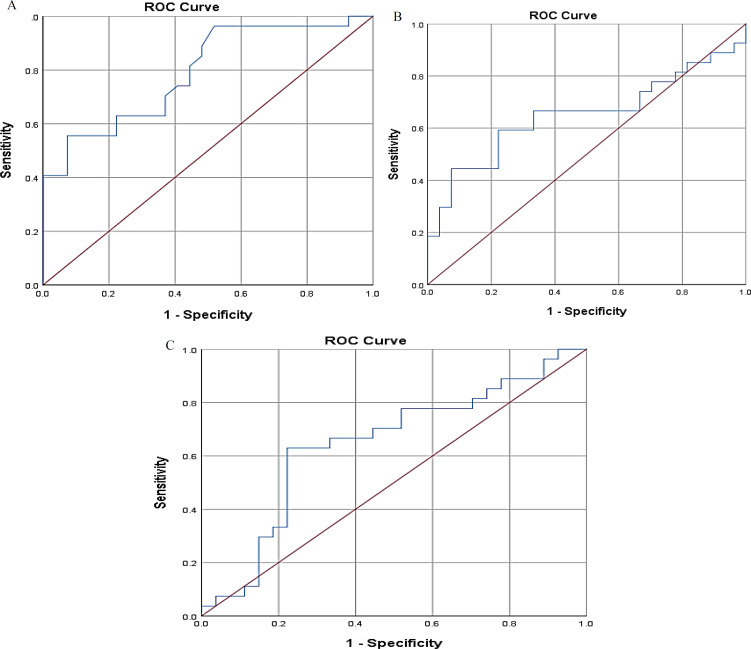
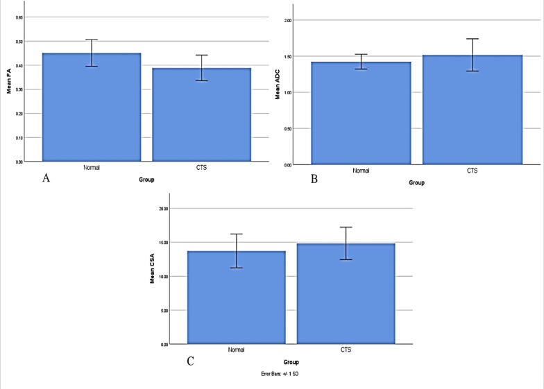
背景:腕管综合征(Carpal tunnel syndrome, CTS)是一种常见的周围神经卡压障碍,通过临床体征和症状诊断,并通过神经传导研究(NCSs)确诊。虽然NCS是一种半侵入性手术,但磁共振成像(MRI)是一种非侵入性诊断工具,可检测宏观神经异常并评估患者的手术或药物治疗方案。本研究通过比较磁共振神经成像(MRN)与电诊断在CTS患者和健康人中的诊断和分级价值。方法:对27例CTS患者腕关节和27例健康腕关节进行横断面研究。经病史和体格检查后,采用NCS确认并确定CTS的严重程度,然后采用MRN和扩散张量成像(DTI)计算表观扩散系数(ADC)、分数各向异性(FA)和横截面积(CSA)。结果:对18例CTS患者(27条正中神经)和15例健康对照(27条正中神经)进行评估。CTS组平均FA显著低于对照组(0.38±0.05 vs. 0.45±0.06,P < 0.001)。CTS患者的平均CSA和ADC较高,但无统计学意义。FA的诊断截止值为0.42,敏感性为70.4%,特异性为63%。结论:mri联合DTI是一种有效的、无创的CTS诊断技术。FA测量显示出足够的敏感性和特异性来区分CTS患者和健康个体。
Assessment of median nerve with magnetic resonance neurography in cases with carpal tunnel syndrome and controls.
Background: Carpal tunnel syndrome (CTS) is a common peripheral nerve entrapment disorder that is diagnosed using clinical signs and symptoms and confirmed via nerve conduction studies (NCSs). While NCS is a semi-invasive procedure, magnetic resonance imaging (MRI) is a non-invasive diagnostic tool that detects macroscopic nerve abnormalities and evaluates a patient's surgical or medication treatment options. This study assessed magnetic resonance neurography (MRN)'s diagnostic and grading value by comparing it to electrodiagnostic studies in patients with CTS and healthy individuals. Methods: This was a cross-sectional study on 27 wrists with CTS and 27 healthy wrists. After history taking and physical examination, we employed an NCS to confirm and determine the severity of CTS, then MRN and diffusion tensor imaging (DTI) were used to calculate apparent diffusion coefficient (ADC), fractional anisotropy (FA), and cross-sectional area (CSA). Results: 18 patients with CTS (27 median nerves) and 15 healthy controls (27 median nerves) were evaluated. The mean FA in the CTS group was significantly lower (0.38 ± 0.05 vs. 0.45 ± 0.06, P < 0.001). The mean CSA and ADC were higher in patients with CTS but not statistically significant. FA's diagnostic cut-off was 0.42, with a sensitivity of 70.4% and a specificity of 63%. Conclusion: MRN with DTI can be an effective and non-invasive diagnostic technique for the detection of CTS. The FA measure demonstrated adequate sensitivity and specificity for differentiating patients with CTS from healthy individuals.


 分享
分享
 求助内容:
求助内容: 应助结果提醒方式:
 扫码关注我们
