{"title":"810 nm二极管激光与光动力治疗减少种植体周围黏膜炎患者微生物菌群的体内比较研究。","authors":"Poonam Siwach, Reshu Sanan, Abhishek Nagpal, Omkar Krishna Shetty, Amit Bhardwaj, Mukesh Sharma","doi":"10.4103/jips.jips_64_24","DOIUrl":null,"url":null,"abstract":"<p><strong>Aim: </strong>The aim of this study was to investigate and compare the antimicrobial effects of an 810-nanometer diode laser, utilizing or not utilizing toluidine blue as a photosensitizer, in the management of peri-implant mucositis.</p><p><strong>Settings and design: </strong>The present study was carried out in 30 implant sites in 15 patients with peri-implant mucositis with a specific inclusion and exclusion criteria. 15 sites were treated utilizing a diode laser (control group) and 15 with photodynamic therapy (test group) in a split-mouth format.</p><p><strong>Materials and methods: </strong>Samples were taken from the gingival sulcus with the help of plastic curettes from control and test sites both at baseline and at 3 months for microbiological analysis.</p><p><strong>Statistical analysis used: </strong>Shapiro-Wilk Test was used to check deviation from normality. Wilcoxon signed-rank test was used to analyse the two dependent groups.</p><p><strong>Results: </strong>Intragroup comparison was found to be statistically significant when compared at baseline and at 3 months in the photodynamic (P = 0.001) and diode laser groups (P = 0.001), respectively. No statistically significant reduction in bacterial count was found at baseline (P = 0.1) and at 3 months (P = 0.5) when the diode laser group and photodynamic group were compared with each other (intergroup).</p><p><strong>Conclusion: </strong>Within the limitations of the study, it can be concluded that there is a definitive reduction in pathogenic bacteria with both interventions and PDT offers clinically visible benefits in the treatment of peri-implant mucositis.</p>","PeriodicalId":22669,"journal":{"name":"The Journal of Indian Prosthodontic Society","volume":"25 1","pages":"40-45"},"PeriodicalIF":1.0000,"publicationDate":"2025-01-01","publicationTypes":"Journal Article","fieldsOfStudy":null,"isOpenAccess":false,"openAccessPdf":"https://www.ncbi.nlm.nih.gov/pmc/articles/PMC11853942/pdf/","citationCount":"0","resultStr":"{\"title\":\"Comparison of the antibacterial efficacy of 810 nm diode laser and photodynamic therapy in reducing microbial flora in patients with peri-implant mucositis - An in vivo study.\",\"authors\":\"Poonam Siwach, Reshu Sanan, Abhishek Nagpal, Omkar Krishna Shetty, Amit Bhardwaj, Mukesh Sharma\",\"doi\":\"10.4103/jips.jips_64_24\",\"DOIUrl\":null,\"url\":null,\"abstract\":\"<p><strong>Aim: </strong>The aim of this study was to investigate and compare the antimicrobial effects of an 810-nanometer diode laser, utilizing or not utilizing toluidine blue as a photosensitizer, in the management of peri-implant mucositis.</p><p><strong>Settings and design: </strong>The present study was carried out in 30 implant sites in 15 patients with peri-implant mucositis with a specific inclusion and exclusion criteria. 15 sites were treated utilizing a diode laser (control group) and 15 with photodynamic therapy (test group) in a split-mouth format.</p><p><strong>Materials and methods: </strong>Samples were taken from the gingival sulcus with the help of plastic curettes from control and test sites both at baseline and at 3 months for microbiological analysis.</p><p><strong>Statistical analysis used: </strong>Shapiro-Wilk Test was used to check deviation from normality. Wilcoxon signed-rank test was used to analyse the two dependent groups.</p><p><strong>Results: </strong>Intragroup comparison was found to be statistically significant when compared at baseline and at 3 months in the photodynamic (P = 0.001) and diode laser groups (P = 0.001), respectively. No statistically significant reduction in bacterial count was found at baseline (P = 0.1) and at 3 months (P = 0.5) when the diode laser group and photodynamic group were compared with each other (intergroup).</p><p><strong>Conclusion: </strong>Within the limitations of the study, it can be concluded that there is a definitive reduction in pathogenic bacteria with both interventions and PDT offers clinically visible benefits in the treatment of peri-implant mucositis.</p>\",\"PeriodicalId\":22669,\"journal\":{\"name\":\"The Journal of Indian Prosthodontic Society\",\"volume\":\"25 1\",\"pages\":\"40-45\"},\"PeriodicalIF\":1.0000,\"publicationDate\":\"2025-01-01\",\"publicationTypes\":\"Journal Article\",\"fieldsOfStudy\":null,\"isOpenAccess\":false,\"openAccessPdf\":\"https://www.ncbi.nlm.nih.gov/pmc/articles/PMC11853942/pdf/\",\"citationCount\":\"0\",\"resultStr\":null,\"platform\":\"Semanticscholar\",\"paperid\":null,\"PeriodicalName\":\"The Journal of Indian Prosthodontic Society\",\"FirstCategoryId\":\"1085\",\"ListUrlMain\":\"https://doi.org/10.4103/jips.jips_64_24\",\"RegionNum\":0,\"RegionCategory\":null,\"ArticlePicture\":[],\"TitleCN\":null,\"AbstractTextCN\":null,\"PMCID\":null,\"EPubDate\":\"2025/1/3 0:00:00\",\"PubModel\":\"Epub\",\"JCR\":\"Q3\",\"JCRName\":\"DENTISTRY, ORAL SURGERY & MEDICINE\",\"Score\":null,\"Total\":0}","platform":"Semanticscholar","paperid":null,"PeriodicalName":"The Journal of Indian Prosthodontic Society","FirstCategoryId":"1085","ListUrlMain":"https://doi.org/10.4103/jips.jips_64_24","RegionNum":0,"RegionCategory":null,"ArticlePicture":[],"TitleCN":null,"AbstractTextCN":null,"PMCID":null,"EPubDate":"2025/1/3 0:00:00","PubModel":"Epub","JCR":"Q3","JCRName":"DENTISTRY, ORAL SURGERY & MEDICINE","Score":null,"Total":0}
Comparison of the antibacterial efficacy of 810 nm diode laser and photodynamic therapy in reducing microbial flora in patients with peri-implant mucositis - An in vivo study.
Aim: The aim of this study was to investigate and compare the antimicrobial effects of an 810-nanometer diode laser, utilizing or not utilizing toluidine blue as a photosensitizer, in the management of peri-implant mucositis.
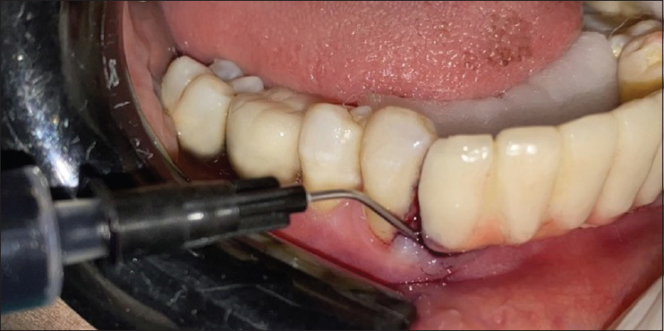
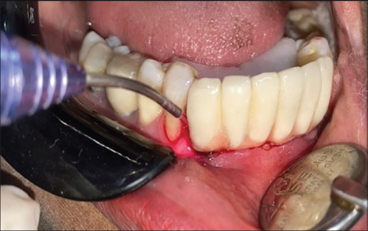
Settings and design: The present study was carried out in 30 implant sites in 15 patients with peri-implant mucositis with a specific inclusion and exclusion criteria. 15 sites were treated utilizing a diode laser (control group) and 15 with photodynamic therapy (test group) in a split-mouth format.
Materials and methods: Samples were taken from the gingival sulcus with the help of plastic curettes from control and test sites both at baseline and at 3 months for microbiological analysis.
Statistical analysis used: Shapiro-Wilk Test was used to check deviation from normality. Wilcoxon signed-rank test was used to analyse the two dependent groups.
Results: Intragroup comparison was found to be statistically significant when compared at baseline and at 3 months in the photodynamic (P = 0.001) and diode laser groups (P = 0.001), respectively. No statistically significant reduction in bacterial count was found at baseline (P = 0.1) and at 3 months (P = 0.5) when the diode laser group and photodynamic group were compared with each other (intergroup).
Conclusion: Within the limitations of the study, it can be concluded that there is a definitive reduction in pathogenic bacteria with both interventions and PDT offers clinically visible benefits in the treatment of peri-implant mucositis.


 分享
分享
 求助内容:
求助内容: 应助结果提醒方式:
 扫码关注我们
