{"title":"铣削和三维打印聚甲基丙烯酸甲酯假体在单冠、前桥和墩基桥中的边缘配合比较:一项体外研究。","authors":"Aman Merchant, Kiran Kumar Pandurangan, Amrutha Dinesh Shenoy, Deepak Nallaswamy, Pooja Nilesh Singh","doi":"10.4103/jips.jips_40_24","DOIUrl":null,"url":null,"abstract":"<p><strong>Aim: </strong>The purpose of this in vitro study was to compare the marginal fit of various three-dimensional (3D) printed and milled polymethylmethacrylate (PMMA) dental prostheses.</p><p><strong>Settings and design: </strong>The study was conducted in an in vitro study setting.</p><p><strong>Materials and methods: </strong>With a sample size of 45 for each fabrication method, this investigation compared the marginal fit of milled (Group 1) and 3D printed (Group 2) PMMA dental prostheses across different designs. The selection of samples was based on G*Power calculations. Tooth preparations were conducted on a typodont jaw set, followed by digital scanning and design processes. Computer-aided design and computer-aided manufacturing milling and 3D printing were employed for the fabrication of prostheses. The assessment of marginal accuracy at specific points was performed using a stereomicroscope.</p><p><strong>Statistical analysis used: </strong>Independent t-tests were used to evaluate marginal discrepancies between milled and printed prosthetic designs at specific tooth surfaces. Univariate analysis of variance assessed marginal discrepancies across prosthesis designs and fabrication methods, with the Tukey post hoc test for significantly different results (α =0.05).</p><p><strong>Results: </strong>Milled single crowns exhibited superior accuracy (61.50 ± 4.852 μ) compared to printed ones (65.74 ± 7.311 μ) (t = -1.868, P = 0.037). Similar trends were observed for other designs, emphasizing the impact of both prosthesis design and fabrication method on marginal fit. Notable discrepancies were found on the distal surfaces of the anterior bridge crossing midline design (t = -3.262, P = 0.003).</p><p><strong>Conclusion: </strong>Milled prostheses exhibited superior marginal fit as compared to 3D printed prostheses, with differences within clinically acceptable limits.</p>","PeriodicalId":22669,"journal":{"name":"The Journal of Indian Prosthodontic Society","volume":"25 1","pages":"67-73"},"PeriodicalIF":1.0000,"publicationDate":"2025-01-01","publicationTypes":"Journal Article","fieldsOfStudy":null,"isOpenAccess":false,"openAccessPdf":"https://www.ncbi.nlm.nih.gov/pmc/articles/PMC11853941/pdf/","citationCount":"0","resultStr":"{\"title\":\"Comparison of marginal fit between milled and three-dimensional printed polymethylmethacrylate prostheses for single crowns, anterior bridges, and pier abutment bridges: An in vitro study.\",\"authors\":\"Aman Merchant, Kiran Kumar Pandurangan, Amrutha Dinesh Shenoy, Deepak Nallaswamy, Pooja Nilesh Singh\",\"doi\":\"10.4103/jips.jips_40_24\",\"DOIUrl\":null,\"url\":null,\"abstract\":\"<p><strong>Aim: </strong>The purpose of this in vitro study was to compare the marginal fit of various three-dimensional (3D) printed and milled polymethylmethacrylate (PMMA) dental prostheses.</p><p><strong>Settings and design: </strong>The study was conducted in an in vitro study setting.</p><p><strong>Materials and methods: </strong>With a sample size of 45 for each fabrication method, this investigation compared the marginal fit of milled (Group 1) and 3D printed (Group 2) PMMA dental prostheses across different designs. The selection of samples was based on G*Power calculations. Tooth preparations were conducted on a typodont jaw set, followed by digital scanning and design processes. Computer-aided design and computer-aided manufacturing milling and 3D printing were employed for the fabrication of prostheses. The assessment of marginal accuracy at specific points was performed using a stereomicroscope.</p><p><strong>Statistical analysis used: </strong>Independent t-tests were used to evaluate marginal discrepancies between milled and printed prosthetic designs at specific tooth surfaces. Univariate analysis of variance assessed marginal discrepancies across prosthesis designs and fabrication methods, with the Tukey post hoc test for significantly different results (α =0.05).</p><p><strong>Results: </strong>Milled single crowns exhibited superior accuracy (61.50 ± 4.852 μ) compared to printed ones (65.74 ± 7.311 μ) (t = -1.868, P = 0.037). Similar trends were observed for other designs, emphasizing the impact of both prosthesis design and fabrication method on marginal fit. Notable discrepancies were found on the distal surfaces of the anterior bridge crossing midline design (t = -3.262, P = 0.003).</p><p><strong>Conclusion: </strong>Milled prostheses exhibited superior marginal fit as compared to 3D printed prostheses, with differences within clinically acceptable limits.</p>\",\"PeriodicalId\":22669,\"journal\":{\"name\":\"The Journal of Indian Prosthodontic Society\",\"volume\":\"25 1\",\"pages\":\"67-73\"},\"PeriodicalIF\":1.0000,\"publicationDate\":\"2025-01-01\",\"publicationTypes\":\"Journal Article\",\"fieldsOfStudy\":null,\"isOpenAccess\":false,\"openAccessPdf\":\"https://www.ncbi.nlm.nih.gov/pmc/articles/PMC11853941/pdf/\",\"citationCount\":\"0\",\"resultStr\":null,\"platform\":\"Semanticscholar\",\"paperid\":null,\"PeriodicalName\":\"The Journal of Indian Prosthodontic Society\",\"FirstCategoryId\":\"1085\",\"ListUrlMain\":\"https://doi.org/10.4103/jips.jips_40_24\",\"RegionNum\":0,\"RegionCategory\":null,\"ArticlePicture\":[],\"TitleCN\":null,\"AbstractTextCN\":null,\"PMCID\":null,\"EPubDate\":\"2025/1/3 0:00:00\",\"PubModel\":\"Epub\",\"JCR\":\"Q3\",\"JCRName\":\"DENTISTRY, ORAL SURGERY & MEDICINE\",\"Score\":null,\"Total\":0}","platform":"Semanticscholar","paperid":null,"PeriodicalName":"The Journal of Indian Prosthodontic Society","FirstCategoryId":"1085","ListUrlMain":"https://doi.org/10.4103/jips.jips_40_24","RegionNum":0,"RegionCategory":null,"ArticlePicture":[],"TitleCN":null,"AbstractTextCN":null,"PMCID":null,"EPubDate":"2025/1/3 0:00:00","PubModel":"Epub","JCR":"Q3","JCRName":"DENTISTRY, ORAL SURGERY & MEDICINE","Score":null,"Total":0}
引用次数: 0
摘要
目的:比较各种三维(3D)打印和研磨的聚甲基丙烯酸甲酯(PMMA)口腔修复体的边缘配合度。环境和设计:本研究在体外研究环境中进行。材料和方法:每种制造方法的样本量为45,本研究比较了不同设计的磨铣(组1)和3D打印(组2)PMMA牙科假体的边际配合。样本的选择基于G*Power计算。牙齿的准备工作进行了排版颌骨设置,其次是数字扫描和设计过程。采用计算机辅助设计和计算机辅助制造,采用铣削和3D打印技术制作假肢。在特定点的边缘精度的评估是使用立体显微镜进行的。采用统计分析:使用独立t检验来评估特定牙齿表面磨铣和打印假体设计之间的边际差异。单因素方差分析评估假体设计和制造方法之间的边际差异,Tukey事后检验结果差异显著(α =0.05)。结果:单冠铣削精度(61.50±4.852 μ)优于打印精度(65.74±7.311 μ) (t = -1.868, P = 0.037)。在其他设计中也观察到类似的趋势,强调了假体设计和制造方法对边缘拟合的影响。在穿过中线设计的前桥远端表面发现显著差异(t = -3.262, P = 0.003)。结论:与3D打印假体相比,磨铣假体具有更好的边缘配合,其差异在临床可接受的范围内。
Comparison of marginal fit between milled and three-dimensional printed polymethylmethacrylate prostheses for single crowns, anterior bridges, and pier abutment bridges: An in vitro study.
Aim: The purpose of this in vitro study was to compare the marginal fit of various three-dimensional (3D) printed and milled polymethylmethacrylate (PMMA) dental prostheses.
Settings and design: The study was conducted in an in vitro study setting.
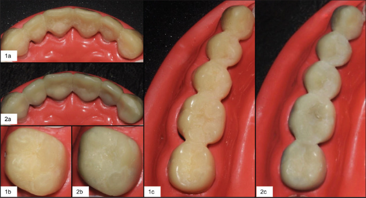
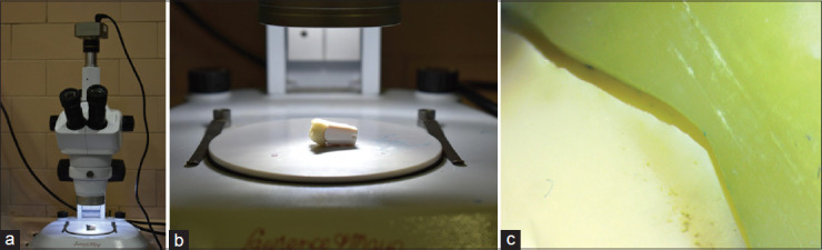
Materials and methods: With a sample size of 45 for each fabrication method, this investigation compared the marginal fit of milled (Group 1) and 3D printed (Group 2) PMMA dental prostheses across different designs. The selection of samples was based on G*Power calculations. Tooth preparations were conducted on a typodont jaw set, followed by digital scanning and design processes. Computer-aided design and computer-aided manufacturing milling and 3D printing were employed for the fabrication of prostheses. The assessment of marginal accuracy at specific points was performed using a stereomicroscope.
Statistical analysis used: Independent t-tests were used to evaluate marginal discrepancies between milled and printed prosthetic designs at specific tooth surfaces. Univariate analysis of variance assessed marginal discrepancies across prosthesis designs and fabrication methods, with the Tukey post hoc test for significantly different results (α =0.05).
Results: Milled single crowns exhibited superior accuracy (61.50 ± 4.852 μ) compared to printed ones (65.74 ± 7.311 μ) (t = -1.868, P = 0.037). Similar trends were observed for other designs, emphasizing the impact of both prosthesis design and fabrication method on marginal fit. Notable discrepancies were found on the distal surfaces of the anterior bridge crossing midline design (t = -3.262, P = 0.003).
Conclusion: Milled prostheses exhibited superior marginal fit as compared to 3D printed prostheses, with differences within clinically acceptable limits.


 分享
分享
 求助内容:
求助内容: 应助结果提醒方式:
 扫码关注我们
