{"title":"自发性脑脊液鼻漏作为特发性颅内高压的主要表现,管理策略和临床结果。","authors":"Ahmed Elshanawany, Farrag Mohammad","doi":"10.25259/SNI_560_2024","DOIUrl":null,"url":null,"abstract":"<p><strong>Background: </strong>Causes of cerebrospinal fluid (CSF) rhinorrhea could be divided into primary (spontaneous) and secondary (head trauma and iatrogenic). Idiopathic intracranial hypertension (IIH) has emerged as a cause for spontaneous CSF rhinorrhea but is still underestimated, may be overlooked and needs special consideration in management. The objective of this study is to demonstrate spontaneous CSF rhinorrhea as the primary presentation of IIH and explore the algorithm of management.</p><p><strong>Methods: </strong>All patients with spontaneous (primary) CSF rhinorrhea were included with complete clinical and radiological assessment. We performed lumbar puncture and CSF pressure measurements in the lateral decubitus position for all included patients to detect those with intracranial hypertension. A pressure of 20 cmH2O in cases of CSF rhinorrhea is considered a cutoff for diagnosing raised intracranial pressure. When intracranial hypertension was diagnosed, patients were subjected immediately to lumboperitoneal shunt. If CSF leakage stopped after shunt insertion, we would not perform skull base repair, and the patient was sent for follow-up. However, if CSF leakage did not stop after shunt insertion despite normalization of intracranial tension or recurrence of CSF rhinorrhea despite shunt patency or there was intracranial pneumocephalus, skull base repair would be performed.</p><p><strong>Results: </strong>During the period of the study, 293 cases of CSF rhinorrhea were seen. Only 42 (14.3%) patients were diagnosed with spontaneous CSF rhinorrhea, and the remaining were posttraumatic. Thirty-seven patients (88.1%) of 42 patients revealed high CSF pressure readings. All 37 patients received lumboperitoneal shunt followed by CSF rhinorrhea stoppage. Later, during follow-up, 7 patients developed recurrence of leakage; 3 of them revealed shunt obstruction, and rhinorrhea improved after shunt revision. The other 4 patients revealed patent shunt and needed skull base repair.</p><p><strong>Conclusion: </strong>Spontaneous CSF rhinorrhea is considered secondary to IIH until proven otherwise. Initial placement of lumboperitoneal shunt may provide an effective alternative to skull base repair for the treatment of patients with IIH presenting with CSF rhinorrhea.</p>","PeriodicalId":94217,"journal":{"name":"Surgical neurology international","volume":"15 ","pages":"458"},"PeriodicalIF":0.0000,"publicationDate":"2024-12-11","publicationTypes":"Journal Article","fieldsOfStudy":null,"isOpenAccess":false,"openAccessPdf":"https://www.ncbi.nlm.nih.gov/pmc/articles/PMC11704438/pdf/","citationCount":"0","resultStr":"{\"title\":\"Spontaneous cerebrospinal fluid rhinorrhea as a primary presentation of idiopathic intracranial hypertension, management strategies, and clinical outcome.\",\"authors\":\"Ahmed Elshanawany, Farrag Mohammad\",\"doi\":\"10.25259/SNI_560_2024\",\"DOIUrl\":null,\"url\":null,\"abstract\":\"<p><strong>Background: </strong>Causes of cerebrospinal fluid (CSF) rhinorrhea could be divided into primary (spontaneous) and secondary (head trauma and iatrogenic). Idiopathic intracranial hypertension (IIH) has emerged as a cause for spontaneous CSF rhinorrhea but is still underestimated, may be overlooked and needs special consideration in management. The objective of this study is to demonstrate spontaneous CSF rhinorrhea as the primary presentation of IIH and explore the algorithm of management.</p><p><strong>Methods: </strong>All patients with spontaneous (primary) CSF rhinorrhea were included with complete clinical and radiological assessment. We performed lumbar puncture and CSF pressure measurements in the lateral decubitus position for all included patients to detect those with intracranial hypertension. A pressure of 20 cmH2O in cases of CSF rhinorrhea is considered a cutoff for diagnosing raised intracranial pressure. When intracranial hypertension was diagnosed, patients were subjected immediately to lumboperitoneal shunt. If CSF leakage stopped after shunt insertion, we would not perform skull base repair, and the patient was sent for follow-up. However, if CSF leakage did not stop after shunt insertion despite normalization of intracranial tension or recurrence of CSF rhinorrhea despite shunt patency or there was intracranial pneumocephalus, skull base repair would be performed.</p><p><strong>Results: </strong>During the period of the study, 293 cases of CSF rhinorrhea were seen. Only 42 (14.3%) patients were diagnosed with spontaneous CSF rhinorrhea, and the remaining were posttraumatic. Thirty-seven patients (88.1%) of 42 patients revealed high CSF pressure readings. All 37 patients received lumboperitoneal shunt followed by CSF rhinorrhea stoppage. Later, during follow-up, 7 patients developed recurrence of leakage; 3 of them revealed shunt obstruction, and rhinorrhea improved after shunt revision. The other 4 patients revealed patent shunt and needed skull base repair.</p><p><strong>Conclusion: </strong>Spontaneous CSF rhinorrhea is considered secondary to IIH until proven otherwise. Initial placement of lumboperitoneal shunt may provide an effective alternative to skull base repair for the treatment of patients with IIH presenting with CSF rhinorrhea.</p>\",\"PeriodicalId\":94217,\"journal\":{\"name\":\"Surgical neurology international\",\"volume\":\"15 \",\"pages\":\"458\"},\"PeriodicalIF\":0.0000,\"publicationDate\":\"2024-12-11\",\"publicationTypes\":\"Journal Article\",\"fieldsOfStudy\":null,\"isOpenAccess\":false,\"openAccessPdf\":\"https://www.ncbi.nlm.nih.gov/pmc/articles/PMC11704438/pdf/\",\"citationCount\":\"0\",\"resultStr\":null,\"platform\":\"Semanticscholar\",\"paperid\":null,\"PeriodicalName\":\"Surgical neurology international\",\"FirstCategoryId\":\"1085\",\"ListUrlMain\":\"https://doi.org/10.25259/SNI_560_2024\",\"RegionNum\":0,\"RegionCategory\":null,\"ArticlePicture\":[],\"TitleCN\":null,\"AbstractTextCN\":null,\"PMCID\":null,\"EPubDate\":\"2024/1/1 0:00:00\",\"PubModel\":\"eCollection\",\"JCR\":\"\",\"JCRName\":\"\",\"Score\":null,\"Total\":0}","platform":"Semanticscholar","paperid":null,"PeriodicalName":"Surgical neurology international","FirstCategoryId":"1085","ListUrlMain":"https://doi.org/10.25259/SNI_560_2024","RegionNum":0,"RegionCategory":null,"ArticlePicture":[],"TitleCN":null,"AbstractTextCN":null,"PMCID":null,"EPubDate":"2024/1/1 0:00:00","PubModel":"eCollection","JCR":"","JCRName":"","Score":null,"Total":0}
Spontaneous cerebrospinal fluid rhinorrhea as a primary presentation of idiopathic intracranial hypertension, management strategies, and clinical outcome.
Background: Causes of cerebrospinal fluid (CSF) rhinorrhea could be divided into primary (spontaneous) and secondary (head trauma and iatrogenic). Idiopathic intracranial hypertension (IIH) has emerged as a cause for spontaneous CSF rhinorrhea but is still underestimated, may be overlooked and needs special consideration in management. The objective of this study is to demonstrate spontaneous CSF rhinorrhea as the primary presentation of IIH and explore the algorithm of management.
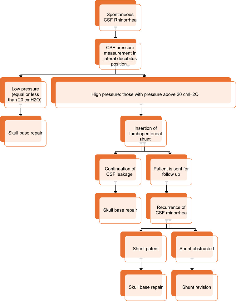
Methods: All patients with spontaneous (primary) CSF rhinorrhea were included with complete clinical and radiological assessment. We performed lumbar puncture and CSF pressure measurements in the lateral decubitus position for all included patients to detect those with intracranial hypertension. A pressure of 20 cmH2O in cases of CSF rhinorrhea is considered a cutoff for diagnosing raised intracranial pressure. When intracranial hypertension was diagnosed, patients were subjected immediately to lumboperitoneal shunt. If CSF leakage stopped after shunt insertion, we would not perform skull base repair, and the patient was sent for follow-up. However, if CSF leakage did not stop after shunt insertion despite normalization of intracranial tension or recurrence of CSF rhinorrhea despite shunt patency or there was intracranial pneumocephalus, skull base repair would be performed.
Results: During the period of the study, 293 cases of CSF rhinorrhea were seen. Only 42 (14.3%) patients were diagnosed with spontaneous CSF rhinorrhea, and the remaining were posttraumatic. Thirty-seven patients (88.1%) of 42 patients revealed high CSF pressure readings. All 37 patients received lumboperitoneal shunt followed by CSF rhinorrhea stoppage. Later, during follow-up, 7 patients developed recurrence of leakage; 3 of them revealed shunt obstruction, and rhinorrhea improved after shunt revision. The other 4 patients revealed patent shunt and needed skull base repair.
Conclusion: Spontaneous CSF rhinorrhea is considered secondary to IIH until proven otherwise. Initial placement of lumboperitoneal shunt may provide an effective alternative to skull base repair for the treatment of patients with IIH presenting with CSF rhinorrhea.


 分享
分享

 求助内容:
求助内容: 应助结果提醒方式:
 扫码关注我们
