Yujiro Ide, Dominik Daniel Gabbert, Jan Hinnerk Hansen, Anselm Uebing, Inga Voges
{"title":"Fontan患者和双心室先天性心脏病患者的肝脏T1定位——静脉充血对弥漫性肝病的影响","authors":"Yujiro Ide, Dominik Daniel Gabbert, Jan Hinnerk Hansen, Anselm Uebing, Inga Voges","doi":"10.1007/s10554-024-03314-5","DOIUrl":null,"url":null,"abstract":"<p><p>T1 relaxation time quantification on parametric maps is routinely used in cardiac imaging and may serve as a non-invasive biomarker for diffuse liver disease. In this study, we aimed to investigate the relationship between liver T1 values and cardiac function in patients with congenital heart disease (CHD) and compared patients with a biventricular circulation (BVC) to those with a Fontan circulation (FC). Magnetic resonance images from patients with CHD, obtained between June and December 2023 on a 1.5 T machine, were retrospectively reviewed. The examinations included cardiac cine sequences to assess ventricular mass and volumes, along with liver T1 mapping. T1 values were measured in eight liver segments and were compared with ventricular mass and volumes in patients with BVC and FC. In total, 104 patients (75 with BVC and 29 with FC) were included. T1 values varied significantly among the eight liver segments in both patient groups. In an age-matched comparison, patients with FC had significantly higher T1 values in all liver segments. In patients with BVC and right ventricular (RV) enlargement, a positive correlation between RV volume and T1 values in the right liver lobe was found (R > 0.504, p < 0.033). In patients with FC, the T1 values did not differ between patients with an extracardiac conduit or a lateral tunnel. Liver T1 mapping suggests more severe liver affection in patients with FC compared to those with BVC. It seems a valuable addition to cardiovascular magnetic resonance for patients who are at risk of systemic venous congestion.</p>","PeriodicalId":94227,"journal":{"name":"The international journal of cardiovascular imaging","volume":" ","pages":"347-358"},"PeriodicalIF":1.5000,"publicationDate":"2025-02-01","publicationTypes":"Journal Article","fieldsOfStudy":null,"isOpenAccess":false,"openAccessPdf":"https://www.ncbi.nlm.nih.gov/pmc/articles/PMC11811443/pdf/","citationCount":"0","resultStr":"{\"title\":\"Liver T1 mapping in Fontan patients and patients with biventricular congenital heart disease - insights into the effects of venous congestions on diffuse liver disease.\",\"authors\":\"Yujiro Ide, Dominik Daniel Gabbert, Jan Hinnerk Hansen, Anselm Uebing, Inga Voges\",\"doi\":\"10.1007/s10554-024-03314-5\",\"DOIUrl\":null,\"url\":null,\"abstract\":\"<p><p>T1 relaxation time quantification on parametric maps is routinely used in cardiac imaging and may serve as a non-invasive biomarker for diffuse liver disease. In this study, we aimed to investigate the relationship between liver T1 values and cardiac function in patients with congenital heart disease (CHD) and compared patients with a biventricular circulation (BVC) to those with a Fontan circulation (FC). Magnetic resonance images from patients with CHD, obtained between June and December 2023 on a 1.5 T machine, were retrospectively reviewed. The examinations included cardiac cine sequences to assess ventricular mass and volumes, along with liver T1 mapping. T1 values were measured in eight liver segments and were compared with ventricular mass and volumes in patients with BVC and FC. In total, 104 patients (75 with BVC and 29 with FC) were included. T1 values varied significantly among the eight liver segments in both patient groups. In an age-matched comparison, patients with FC had significantly higher T1 values in all liver segments. In patients with BVC and right ventricular (RV) enlargement, a positive correlation between RV volume and T1 values in the right liver lobe was found (R > 0.504, p < 0.033). In patients with FC, the T1 values did not differ between patients with an extracardiac conduit or a lateral tunnel. Liver T1 mapping suggests more severe liver affection in patients with FC compared to those with BVC. It seems a valuable addition to cardiovascular magnetic resonance for patients who are at risk of systemic venous congestion.</p>\",\"PeriodicalId\":94227,\"journal\":{\"name\":\"The international journal of cardiovascular imaging\",\"volume\":\" \",\"pages\":\"347-358\"},\"PeriodicalIF\":1.5000,\"publicationDate\":\"2025-02-01\",\"publicationTypes\":\"Journal Article\",\"fieldsOfStudy\":null,\"isOpenAccess\":false,\"openAccessPdf\":\"https://www.ncbi.nlm.nih.gov/pmc/articles/PMC11811443/pdf/\",\"citationCount\":\"0\",\"resultStr\":null,\"platform\":\"Semanticscholar\",\"paperid\":null,\"PeriodicalName\":\"The international journal of cardiovascular imaging\",\"FirstCategoryId\":\"1085\",\"ListUrlMain\":\"https://doi.org/10.1007/s10554-024-03314-5\",\"RegionNum\":0,\"RegionCategory\":null,\"ArticlePicture\":[],\"TitleCN\":null,\"AbstractTextCN\":null,\"PMCID\":null,\"EPubDate\":\"2025/1/8 0:00:00\",\"PubModel\":\"Epub\",\"JCR\":\"\",\"JCRName\":\"\",\"Score\":null,\"Total\":0}","platform":"Semanticscholar","paperid":null,"PeriodicalName":"The international journal of cardiovascular imaging","FirstCategoryId":"1085","ListUrlMain":"https://doi.org/10.1007/s10554-024-03314-5","RegionNum":0,"RegionCategory":null,"ArticlePicture":[],"TitleCN":null,"AbstractTextCN":null,"PMCID":null,"EPubDate":"2025/1/8 0:00:00","PubModel":"Epub","JCR":"","JCRName":"","Score":null,"Total":0}
引用次数: 0
摘要
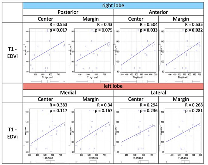
参数图上的T1松弛时间量化通常用于心脏成像,并可作为弥漫性肝脏疾病的非侵入性生物标志物。在这项研究中,我们旨在探讨先天性心脏病(CHD)患者肝脏T1值与心功能的关系,并比较双心室循环(BVC)患者和Fontan循环(FC)患者。回顾性回顾了2023年6月至12月在1.5 T机器上获得的冠心病患者的磁共振图像。检查包括心脏影像序列以评估心室质量和体积,以及肝脏T1制图。测量8个肝节段的T1值,并与BVC和FC患者的心室质量和体积进行比较。共纳入104例患者(75例BVC, 29例FC)。两组患者8个肝段T1值差异显著。在年龄匹配的比较中,FC患者在所有肝段的T1值明显更高。在BVC合并右心室增大的患者中,右心室体积与右肝叶T1值呈正相关(r> 0.504, p
Liver T1 mapping in Fontan patients and patients with biventricular congenital heart disease - insights into the effects of venous congestions on diffuse liver disease.
T1 relaxation time quantification on parametric maps is routinely used in cardiac imaging and may serve as a non-invasive biomarker for diffuse liver disease. In this study, we aimed to investigate the relationship between liver T1 values and cardiac function in patients with congenital heart disease (CHD) and compared patients with a biventricular circulation (BVC) to those with a Fontan circulation (FC). Magnetic resonance images from patients with CHD, obtained between June and December 2023 on a 1.5 T machine, were retrospectively reviewed. The examinations included cardiac cine sequences to assess ventricular mass and volumes, along with liver T1 mapping. T1 values were measured in eight liver segments and were compared with ventricular mass and volumes in patients with BVC and FC. In total, 104 patients (75 with BVC and 29 with FC) were included. T1 values varied significantly among the eight liver segments in both patient groups. In an age-matched comparison, patients with FC had significantly higher T1 values in all liver segments. In patients with BVC and right ventricular (RV) enlargement, a positive correlation between RV volume and T1 values in the right liver lobe was found (R > 0.504, p < 0.033). In patients with FC, the T1 values did not differ between patients with an extracardiac conduit or a lateral tunnel. Liver T1 mapping suggests more severe liver affection in patients with FC compared to those with BVC. It seems a valuable addition to cardiovascular magnetic resonance for patients who are at risk of systemic venous congestion.


 分享
分享

 求助内容:
求助内容: 应助结果提醒方式:
 扫码关注我们
