{"title":"冠突松质骨瘤:文献回顾及罕见病例报告。","authors":"Zygimantas Petronis, Audra Janovskiene, Marijus Leketas","doi":"10.22514/jofph.2024.021","DOIUrl":null,"url":null,"abstract":"<p><p>Osteoma is a rare benign tumor primarily affecting the craniofacial skeleton. Coronary osteomas in the coronoid process are uncommon and asymptomatic until they affect mandibular function. This report presents a case of coronoid osteoma with its diagnosis, treatment and surgical approach. Osteoma is a benign tumor composed of well-differentiated bone tissue, with different origins: central, peripheral and extraskeletal. Mandibular coronoid osteomas are rare but important to consider in symptomatic patients. Mandibular osteoma frequency ranges from 22.8% to 81.3%, with a reported frequency of 15.28% in the maxilla. A 63-year-old female presented with facial deformation, limited mouth opening, and associated symptoms, such as intermittent dizziness and nasal congestion. Imaging revealed a well-defined radiopaque lesion in the coronoid process, displacing surrounding structures. Diagnosis confirmed coronoid osteoma. Surgical removal resulted in satisfactory recovery and improved mouth opening. Coronoid osteomas are rare, with limited reported cases. Osteomas are more prevalent in the mandible body than in other locations. Radiographic imaging and histopathological examination are crucial for diagnosis. Radiographic and histological features distinguish osteomas from other lesions. Etiology remains uncertain, with trauma, temporal muscle hyperactivity, or post-traumatic fibrosis as potential causes. Differential diagnosis involves distinguishing osteoma from osteochondroma, osteoblastoma, exostoses and osteosarcoma. Continued research and reporting are necessary to enhance understanding and management of this rare condition.</p>","PeriodicalId":48800,"journal":{"name":"Journal of Oral & Facial Pain and Headache","volume":"38 2","pages":"126-130"},"PeriodicalIF":2.4000,"publicationDate":"2024-06-01","publicationTypes":"Journal Article","fieldsOfStudy":null,"isOpenAccess":false,"openAccessPdf":"https://www.ncbi.nlm.nih.gov/pmc/articles/PMC11810653/pdf/","citationCount":"0","resultStr":"{\"title\":\"Cancellous osteoma of the coronoid process: a literature review and rare case report.\",\"authors\":\"Zygimantas Petronis, Audra Janovskiene, Marijus Leketas\",\"doi\":\"10.22514/jofph.2024.021\",\"DOIUrl\":null,\"url\":null,\"abstract\":\"<p><p>Osteoma is a rare benign tumor primarily affecting the craniofacial skeleton. Coronary osteomas in the coronoid process are uncommon and asymptomatic until they affect mandibular function. This report presents a case of coronoid osteoma with its diagnosis, treatment and surgical approach. Osteoma is a benign tumor composed of well-differentiated bone tissue, with different origins: central, peripheral and extraskeletal. Mandibular coronoid osteomas are rare but important to consider in symptomatic patients. Mandibular osteoma frequency ranges from 22.8% to 81.3%, with a reported frequency of 15.28% in the maxilla. A 63-year-old female presented with facial deformation, limited mouth opening, and associated symptoms, such as intermittent dizziness and nasal congestion. Imaging revealed a well-defined radiopaque lesion in the coronoid process, displacing surrounding structures. Diagnosis confirmed coronoid osteoma. Surgical removal resulted in satisfactory recovery and improved mouth opening. Coronoid osteomas are rare, with limited reported cases. Osteomas are more prevalent in the mandible body than in other locations. Radiographic imaging and histopathological examination are crucial for diagnosis. Radiographic and histological features distinguish osteomas from other lesions. Etiology remains uncertain, with trauma, temporal muscle hyperactivity, or post-traumatic fibrosis as potential causes. Differential diagnosis involves distinguishing osteoma from osteochondroma, osteoblastoma, exostoses and osteosarcoma. Continued research and reporting are necessary to enhance understanding and management of this rare condition.</p>\",\"PeriodicalId\":48800,\"journal\":{\"name\":\"Journal of Oral & Facial Pain and Headache\",\"volume\":\"38 2\",\"pages\":\"126-130\"},\"PeriodicalIF\":2.4000,\"publicationDate\":\"2024-06-01\",\"publicationTypes\":\"Journal Article\",\"fieldsOfStudy\":null,\"isOpenAccess\":false,\"openAccessPdf\":\"https://www.ncbi.nlm.nih.gov/pmc/articles/PMC11810653/pdf/\",\"citationCount\":\"0\",\"resultStr\":null,\"platform\":\"Semanticscholar\",\"paperid\":null,\"PeriodicalName\":\"Journal of Oral & Facial Pain and Headache\",\"FirstCategoryId\":\"3\",\"ListUrlMain\":\"https://doi.org/10.22514/jofph.2024.021\",\"RegionNum\":3,\"RegionCategory\":\"医学\",\"ArticlePicture\":[],\"TitleCN\":null,\"AbstractTextCN\":null,\"PMCID\":null,\"EPubDate\":\"2024/6/12 0:00:00\",\"PubModel\":\"Epub\",\"JCR\":\"Q2\",\"JCRName\":\"DENTISTRY, ORAL SURGERY & MEDICINE\",\"Score\":null,\"Total\":0}","platform":"Semanticscholar","paperid":null,"PeriodicalName":"Journal of Oral & Facial Pain and Headache","FirstCategoryId":"3","ListUrlMain":"https://doi.org/10.22514/jofph.2024.021","RegionNum":3,"RegionCategory":"医学","ArticlePicture":[],"TitleCN":null,"AbstractTextCN":null,"PMCID":null,"EPubDate":"2024/6/12 0:00:00","PubModel":"Epub","JCR":"Q2","JCRName":"DENTISTRY, ORAL SURGERY & MEDICINE","Score":null,"Total":0}
Cancellous osteoma of the coronoid process: a literature review and rare case report.
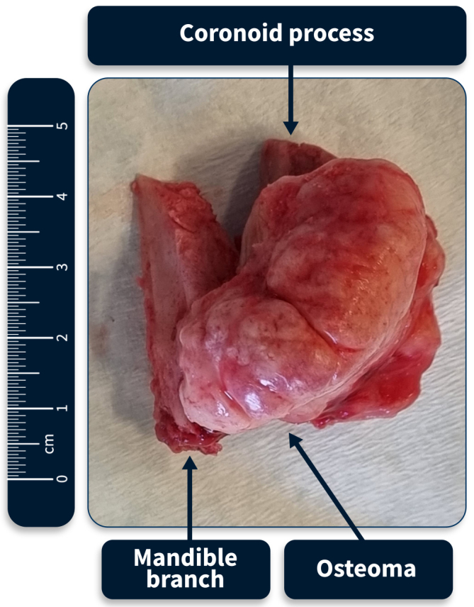
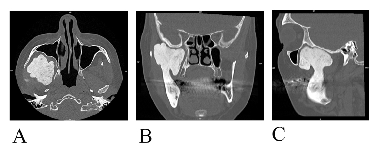
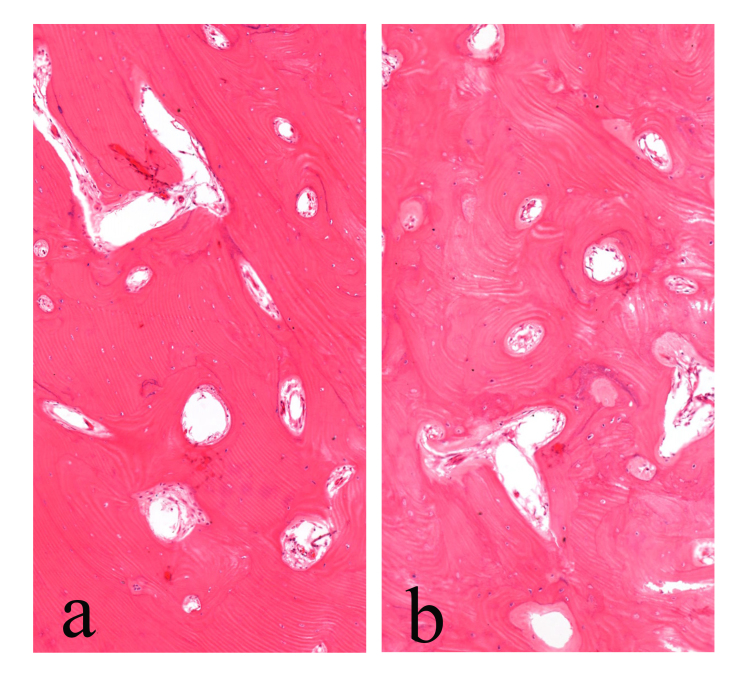
Osteoma is a rare benign tumor primarily affecting the craniofacial skeleton. Coronary osteomas in the coronoid process are uncommon and asymptomatic until they affect mandibular function. This report presents a case of coronoid osteoma with its diagnosis, treatment and surgical approach. Osteoma is a benign tumor composed of well-differentiated bone tissue, with different origins: central, peripheral and extraskeletal. Mandibular coronoid osteomas are rare but important to consider in symptomatic patients. Mandibular osteoma frequency ranges from 22.8% to 81.3%, with a reported frequency of 15.28% in the maxilla. A 63-year-old female presented with facial deformation, limited mouth opening, and associated symptoms, such as intermittent dizziness and nasal congestion. Imaging revealed a well-defined radiopaque lesion in the coronoid process, displacing surrounding structures. Diagnosis confirmed coronoid osteoma. Surgical removal resulted in satisfactory recovery and improved mouth opening. Coronoid osteomas are rare, with limited reported cases. Osteomas are more prevalent in the mandible body than in other locations. Radiographic imaging and histopathological examination are crucial for diagnosis. Radiographic and histological features distinguish osteomas from other lesions. Etiology remains uncertain, with trauma, temporal muscle hyperactivity, or post-traumatic fibrosis as potential causes. Differential diagnosis involves distinguishing osteoma from osteochondroma, osteoblastoma, exostoses and osteosarcoma. Continued research and reporting are necessary to enhance understanding and management of this rare condition.
期刊介绍:
Founded upon sound scientific principles, this journal continues to make important contributions that strongly influence the work of dental and medical professionals involved in treating oral and facial pain, including temporomandibular disorders, and headache. In addition to providing timely scientific research and clinical articles, the journal presents diagnostic techniques and treatment therapies for oral and facial pain, headache, mandibular dysfunction, and occlusion and covers pharmacology, physical therapy, surgery, and other pain-management methods.


 分享
分享
 求助内容:
求助内容: 应助结果提醒方式:
 扫码关注我们
