{"title":"评估胸部CT在肝内胆管癌患者随访中的价值。","authors":"Moamen Abdelaal, Mahmoud Diab, Maguy Farhat, Milind Javle, Mostafa Shehata, Emad Singer, Yomna Khamis, Manal Hassan, Khaled M Elsayes, Janio Szklaruk","doi":"10.21037/jgo-24-365","DOIUrl":null,"url":null,"abstract":"<p><strong>Background: </strong>Following tumor resection, imaging recommendation for the follow-up of patients with intrahepatic cholangiocarcinoma (IHCCA) include frequent chest, abdomen and pelvis computed tomography (CT) imaging. The appropriateness of additional imaging studies is usually derived from their clinical utility. The purpose of this work is to determine the value of chest CT imaging in the follow-up of patients with IHCCA.</p><p><strong>Methods: </strong>Data review of radiology reports of baseline post-operative chest, abdominal, and pelvic CT imaging reports following the resection of IHCCA, and of subsequent follow-up exams. The radiology findings were stratified as intrathoracic metastasis only, combined intra-thoracic and intraabdominal metastasis, intra-abdominal metastases without intrathoracic involvement. We assessed the prevalence of intra-thoracic disease progression in comparison to other groups. Descriptive statistical analysis was carried out using John's Macintosh Project (JMP) statistical software.</p><p><strong>Results: </strong>Eighty-seven patients were included in the study, 6 patients were found to have disease progression in the chest without corresponding disease progression in the abdomen on follow-up CT, accounting for 6.9% of the total. Only four patients had disease progression in the chest with a normal CT chest at baseline (4.6%).</p><p><strong>Conclusions: </strong>The use of short-interval chest CT for surveillance in IHCCA has limited utility, particularly in patients with disease-free abdominal studies.</p>","PeriodicalId":15841,"journal":{"name":"Journal of gastrointestinal oncology","volume":"15 6","pages":"2656-2662"},"PeriodicalIF":2.0000,"publicationDate":"2024-12-31","publicationTypes":"Journal Article","fieldsOfStudy":null,"isOpenAccess":false,"openAccessPdf":"https://www.ncbi.nlm.nih.gov/pmc/articles/PMC11732355/pdf/","citationCount":"0","resultStr":"{\"title\":\"Assessing the value of computed tomography (CT) of the chest in the follow-up of patients with intrahepatic cholangiocarcinoma.\",\"authors\":\"Moamen Abdelaal, Mahmoud Diab, Maguy Farhat, Milind Javle, Mostafa Shehata, Emad Singer, Yomna Khamis, Manal Hassan, Khaled M Elsayes, Janio Szklaruk\",\"doi\":\"10.21037/jgo-24-365\",\"DOIUrl\":null,\"url\":null,\"abstract\":\"<p><strong>Background: </strong>Following tumor resection, imaging recommendation for the follow-up of patients with intrahepatic cholangiocarcinoma (IHCCA) include frequent chest, abdomen and pelvis computed tomography (CT) imaging. The appropriateness of additional imaging studies is usually derived from their clinical utility. The purpose of this work is to determine the value of chest CT imaging in the follow-up of patients with IHCCA.</p><p><strong>Methods: </strong>Data review of radiology reports of baseline post-operative chest, abdominal, and pelvic CT imaging reports following the resection of IHCCA, and of subsequent follow-up exams. The radiology findings were stratified as intrathoracic metastasis only, combined intra-thoracic and intraabdominal metastasis, intra-abdominal metastases without intrathoracic involvement. We assessed the prevalence of intra-thoracic disease progression in comparison to other groups. Descriptive statistical analysis was carried out using John's Macintosh Project (JMP) statistical software.</p><p><strong>Results: </strong>Eighty-seven patients were included in the study, 6 patients were found to have disease progression in the chest without corresponding disease progression in the abdomen on follow-up CT, accounting for 6.9% of the total. Only four patients had disease progression in the chest with a normal CT chest at baseline (4.6%).</p><p><strong>Conclusions: </strong>The use of short-interval chest CT for surveillance in IHCCA has limited utility, particularly in patients with disease-free abdominal studies.</p>\",\"PeriodicalId\":15841,\"journal\":{\"name\":\"Journal of gastrointestinal oncology\",\"volume\":\"15 6\",\"pages\":\"2656-2662\"},\"PeriodicalIF\":2.0000,\"publicationDate\":\"2024-12-31\",\"publicationTypes\":\"Journal Article\",\"fieldsOfStudy\":null,\"isOpenAccess\":false,\"openAccessPdf\":\"https://www.ncbi.nlm.nih.gov/pmc/articles/PMC11732355/pdf/\",\"citationCount\":\"0\",\"resultStr\":null,\"platform\":\"Semanticscholar\",\"paperid\":null,\"PeriodicalName\":\"Journal of gastrointestinal oncology\",\"FirstCategoryId\":\"3\",\"ListUrlMain\":\"https://doi.org/10.21037/jgo-24-365\",\"RegionNum\":4,\"RegionCategory\":\"医学\",\"ArticlePicture\":[],\"TitleCN\":null,\"AbstractTextCN\":null,\"PMCID\":null,\"EPubDate\":\"2024/12/28 0:00:00\",\"PubModel\":\"Epub\",\"JCR\":\"Q3\",\"JCRName\":\"GASTROENTEROLOGY & HEPATOLOGY\",\"Score\":null,\"Total\":0}","platform":"Semanticscholar","paperid":null,"PeriodicalName":"Journal of gastrointestinal oncology","FirstCategoryId":"3","ListUrlMain":"https://doi.org/10.21037/jgo-24-365","RegionNum":4,"RegionCategory":"医学","ArticlePicture":[],"TitleCN":null,"AbstractTextCN":null,"PMCID":null,"EPubDate":"2024/12/28 0:00:00","PubModel":"Epub","JCR":"Q3","JCRName":"GASTROENTEROLOGY & HEPATOLOGY","Score":null,"Total":0}
引用次数: 0
摘要
背景:肿瘤切除后,肝内胆管癌(IHCCA)患者随访的影像学建议包括频繁的胸部、腹部和骨盆CT成像。额外影像学检查的适当性通常源于其临床应用。本工作的目的是确定胸部CT成像在IHCCA患者随访中的价值。方法:回顾IHCCA切除术后胸部、腹部和骨盆基线CT影像学报告以及随后随访检查的资料。影像学表现分为单胸转移、胸腹合并转移、不累及胸内转移。与其他组相比,我们评估了胸内疾病进展的患病率。采用John’s Macintosh Project (JMP)统计软件进行描述性统计分析。结果:纳入研究的87例患者中,随访CT发现6例患者胸部有疾病进展,腹部无相应疾病进展,占总数的6.9%。只有4例患者在基线时胸部CT显示正常,但胸部疾病进展(4.6%)。结论:在IHCCA中使用短间隔胸部CT进行监测的效用有限,特别是在无疾病腹部研究的患者中。
Assessing the value of computed tomography (CT) of the chest in the follow-up of patients with intrahepatic cholangiocarcinoma.
Background: Following tumor resection, imaging recommendation for the follow-up of patients with intrahepatic cholangiocarcinoma (IHCCA) include frequent chest, abdomen and pelvis computed tomography (CT) imaging. The appropriateness of additional imaging studies is usually derived from their clinical utility. The purpose of this work is to determine the value of chest CT imaging in the follow-up of patients with IHCCA.
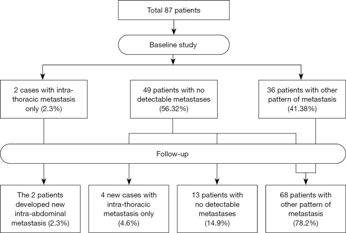
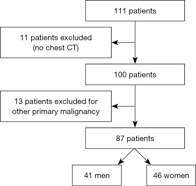
Methods: Data review of radiology reports of baseline post-operative chest, abdominal, and pelvic CT imaging reports following the resection of IHCCA, and of subsequent follow-up exams. The radiology findings were stratified as intrathoracic metastasis only, combined intra-thoracic and intraabdominal metastasis, intra-abdominal metastases without intrathoracic involvement. We assessed the prevalence of intra-thoracic disease progression in comparison to other groups. Descriptive statistical analysis was carried out using John's Macintosh Project (JMP) statistical software.
Results: Eighty-seven patients were included in the study, 6 patients were found to have disease progression in the chest without corresponding disease progression in the abdomen on follow-up CT, accounting for 6.9% of the total. Only four patients had disease progression in the chest with a normal CT chest at baseline (4.6%).
Conclusions: The use of short-interval chest CT for surveillance in IHCCA has limited utility, particularly in patients with disease-free abdominal studies.
期刊介绍:
ournal of Gastrointestinal Oncology (Print ISSN 2078-6891; Online ISSN 2219-679X; J Gastrointest Oncol; JGO), the official journal of Society for Gastrointestinal Oncology (SGO), is an open-access, international peer-reviewed journal. It is published quarterly (Sep. 2010- Dec. 2013), bimonthly (Feb. 2014 -) and openly distributed worldwide.
JGO publishes manuscripts that focus on updated and practical information about diagnosis, prevention and clinical investigations of gastrointestinal cancer treatment. Specific areas of interest include, but not limited to, multimodality therapy, markers, imaging and tumor biology.


 分享
分享
 求助内容:
求助内容: 应助结果提醒方式:
 扫码关注我们
