Mélodie Cyr, Naim Chabaytah, Joud Babik, Behnaz Behmand, Guillaume St-Jean, Shirin A Enger
{"title":"建立规范的小鼠原位直肠内模型用于研究结直肠癌。","authors":"Mélodie Cyr, Naim Chabaytah, Joud Babik, Behnaz Behmand, Guillaume St-Jean, Shirin A Enger","doi":"10.21037/jgo-24-515","DOIUrl":null,"url":null,"abstract":"<p><strong>Background: </strong>Orthotopic models offer a more accurate representation of colorectal cancer (CRC) compared to subcutaneous models. Despite promising results from the reported intra-rectal models, establishing a standardized method for CRC research remains challenging due to model variability, hindering comprehensive studies on CRC pathogenesis and treatment modalities, such as brachytherapy. This study aimed to establish a standardized workflow for an orthotopic intra-rectal animal model to induce the growth of colorectal adenocarcinoma in male and female mice.</p><p><strong>Methods: </strong>HT-29 colorectal adenocarcinoma cells were injected into the rectal mucosa of female (n=21) and male (n=26) non-obese diabetic severe combined immunodeficiency (NOD SCID) gamma (NSG) mice. Mice were placed on a 45° wedge elevating their pelvis for better visualization of the anus. Tumor growth and localization were monitored using a 7-T magnetic resonance imaging (MRI) scanner with rapid acquisition with relaxation echo (RARE) sequence at weeks 1, 2, and 3 post-cell instillation. Once tumors reached 5-8 mm in diameter, the mice were euthanized. Histopathology and immunohistochemical analyses confirmed the tumors' morphology, including necrosis, vascularity (CD-31) and apoptosis (cleaved caspase-3).</p><p><strong>Results: </strong>There was a 92% and 95% tumor growth success rate in male and female mice, respectively. Tumors grew to 5-8 mm in diameter within ~20 days. No significant difference in tumor size was observed between genders. Tumor morphology was consistent across cases. Most tumors exhibited a lack of central blood vessels, accompanied by varying degrees of necrosis and apoptosis, whereas external portions were highly vascularized.</p><p><strong>Conclusions: </strong>An orthotopic intra-rectal model was successfully developed. This model will be used in future studies to evaluate the efficacy of CRC treatments.</p>","PeriodicalId":15841,"journal":{"name":"Journal of gastrointestinal oncology","volume":"15 6","pages":"2578-2587"},"PeriodicalIF":2.1000,"publicationDate":"2024-12-31","publicationTypes":"Journal Article","fieldsOfStudy":null,"isOpenAccess":false,"openAccessPdf":"https://www.ncbi.nlm.nih.gov/pmc/articles/PMC11732354/pdf/","citationCount":"0","resultStr":"{\"title\":\"Establishing a standardized murine orthotopic intra-rectal model for the study of colorectal adenocarcinoma.\",\"authors\":\"Mélodie Cyr, Naim Chabaytah, Joud Babik, Behnaz Behmand, Guillaume St-Jean, Shirin A Enger\",\"doi\":\"10.21037/jgo-24-515\",\"DOIUrl\":null,\"url\":null,\"abstract\":\"<p><strong>Background: </strong>Orthotopic models offer a more accurate representation of colorectal cancer (CRC) compared to subcutaneous models. Despite promising results from the reported intra-rectal models, establishing a standardized method for CRC research remains challenging due to model variability, hindering comprehensive studies on CRC pathogenesis and treatment modalities, such as brachytherapy. This study aimed to establish a standardized workflow for an orthotopic intra-rectal animal model to induce the growth of colorectal adenocarcinoma in male and female mice.</p><p><strong>Methods: </strong>HT-29 colorectal adenocarcinoma cells were injected into the rectal mucosa of female (n=21) and male (n=26) non-obese diabetic severe combined immunodeficiency (NOD SCID) gamma (NSG) mice. Mice were placed on a 45° wedge elevating their pelvis for better visualization of the anus. Tumor growth and localization were monitored using a 7-T magnetic resonance imaging (MRI) scanner with rapid acquisition with relaxation echo (RARE) sequence at weeks 1, 2, and 3 post-cell instillation. Once tumors reached 5-8 mm in diameter, the mice were euthanized. Histopathology and immunohistochemical analyses confirmed the tumors' morphology, including necrosis, vascularity (CD-31) and apoptosis (cleaved caspase-3).</p><p><strong>Results: </strong>There was a 92% and 95% tumor growth success rate in male and female mice, respectively. Tumors grew to 5-8 mm in diameter within ~20 days. No significant difference in tumor size was observed between genders. Tumor morphology was consistent across cases. Most tumors exhibited a lack of central blood vessels, accompanied by varying degrees of necrosis and apoptosis, whereas external portions were highly vascularized.</p><p><strong>Conclusions: </strong>An orthotopic intra-rectal model was successfully developed. This model will be used in future studies to evaluate the efficacy of CRC treatments.</p>\",\"PeriodicalId\":15841,\"journal\":{\"name\":\"Journal of gastrointestinal oncology\",\"volume\":\"15 6\",\"pages\":\"2578-2587\"},\"PeriodicalIF\":2.1000,\"publicationDate\":\"2024-12-31\",\"publicationTypes\":\"Journal Article\",\"fieldsOfStudy\":null,\"isOpenAccess\":false,\"openAccessPdf\":\"https://www.ncbi.nlm.nih.gov/pmc/articles/PMC11732354/pdf/\",\"citationCount\":\"0\",\"resultStr\":null,\"platform\":\"Semanticscholar\",\"paperid\":null,\"PeriodicalName\":\"Journal of gastrointestinal oncology\",\"FirstCategoryId\":\"3\",\"ListUrlMain\":\"https://doi.org/10.21037/jgo-24-515\",\"RegionNum\":4,\"RegionCategory\":\"医学\",\"ArticlePicture\":[],\"TitleCN\":null,\"AbstractTextCN\":null,\"PMCID\":null,\"EPubDate\":\"2024/12/9 0:00:00\",\"PubModel\":\"Epub\",\"JCR\":\"Q3\",\"JCRName\":\"GASTROENTEROLOGY & HEPATOLOGY\",\"Score\":null,\"Total\":0}","platform":"Semanticscholar","paperid":null,"PeriodicalName":"Journal of gastrointestinal oncology","FirstCategoryId":"3","ListUrlMain":"https://doi.org/10.21037/jgo-24-515","RegionNum":4,"RegionCategory":"医学","ArticlePicture":[],"TitleCN":null,"AbstractTextCN":null,"PMCID":null,"EPubDate":"2024/12/9 0:00:00","PubModel":"Epub","JCR":"Q3","JCRName":"GASTROENTEROLOGY & HEPATOLOGY","Score":null,"Total":0}
Establishing a standardized murine orthotopic intra-rectal model for the study of colorectal adenocarcinoma.
Background: Orthotopic models offer a more accurate representation of colorectal cancer (CRC) compared to subcutaneous models. Despite promising results from the reported intra-rectal models, establishing a standardized method for CRC research remains challenging due to model variability, hindering comprehensive studies on CRC pathogenesis and treatment modalities, such as brachytherapy. This study aimed to establish a standardized workflow for an orthotopic intra-rectal animal model to induce the growth of colorectal adenocarcinoma in male and female mice.
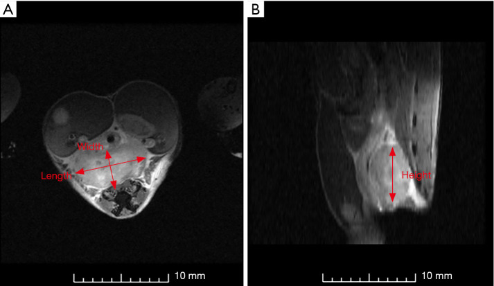
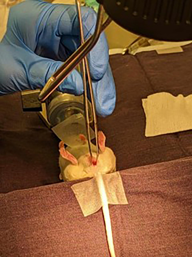
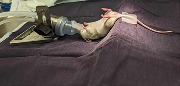
Methods: HT-29 colorectal adenocarcinoma cells were injected into the rectal mucosa of female (n=21) and male (n=26) non-obese diabetic severe combined immunodeficiency (NOD SCID) gamma (NSG) mice. Mice were placed on a 45° wedge elevating their pelvis for better visualization of the anus. Tumor growth and localization were monitored using a 7-T magnetic resonance imaging (MRI) scanner with rapid acquisition with relaxation echo (RARE) sequence at weeks 1, 2, and 3 post-cell instillation. Once tumors reached 5-8 mm in diameter, the mice were euthanized. Histopathology and immunohistochemical analyses confirmed the tumors' morphology, including necrosis, vascularity (CD-31) and apoptosis (cleaved caspase-3).
Results: There was a 92% and 95% tumor growth success rate in male and female mice, respectively. Tumors grew to 5-8 mm in diameter within ~20 days. No significant difference in tumor size was observed between genders. Tumor morphology was consistent across cases. Most tumors exhibited a lack of central blood vessels, accompanied by varying degrees of necrosis and apoptosis, whereas external portions were highly vascularized.
Conclusions: An orthotopic intra-rectal model was successfully developed. This model will be used in future studies to evaluate the efficacy of CRC treatments.
期刊介绍:
ournal of Gastrointestinal Oncology (Print ISSN 2078-6891; Online ISSN 2219-679X; J Gastrointest Oncol; JGO), the official journal of Society for Gastrointestinal Oncology (SGO), is an open-access, international peer-reviewed journal. It is published quarterly (Sep. 2010- Dec. 2013), bimonthly (Feb. 2014 -) and openly distributed worldwide.
JGO publishes manuscripts that focus on updated and practical information about diagnosis, prevention and clinical investigations of gastrointestinal cancer treatment. Specific areas of interest include, but not limited to, multimodality therapy, markers, imaging and tumor biology.


 分享
分享
 求助内容:
求助内容: 应助结果提醒方式:
 扫码关注我们
