Kazuhiro Kato, Hironobu Yasui, Hideo Sato-Akaba, Miho C Emoto, Hirotada G Fujii, Maciej M Kmiec, Periannan Kuppusamy, Masaki Nagane, Tadashi Yamashita, Osamu Inanami
{"title":"无创电子顺磁共振成像检测由铁下垂引起的肿瘤氧化还原失衡。","authors":"Kazuhiro Kato, Hironobu Yasui, Hideo Sato-Akaba, Miho C Emoto, Hirotada G Fujii, Maciej M Kmiec, Periannan Kuppusamy, Masaki Nagane, Tadashi Yamashita, Osamu Inanami","doi":"10.1080/13510002.2025.2454887","DOIUrl":null,"url":null,"abstract":"<p><p>Targeting ferroptosis, cell death caused by the iron-dependent accumulation of lipid peroxides, and disruption of the redox balance are promising strategies in cancer therapy owing to the physiological characteristics of cancer cells. However, the detection of ferroptosis using <i>in vivo</i> imaging remains challenging. We previously reported that redox maps showing the reduction power per unit time of implanted tumor tissues via non-invasive redox imaging using a novel, compact, and portable electron paramagnetic resonance imaging (EPRI) device could be compared with tumor tissue sections. This study aimed to apply the EPRI technique to the <i>in vivo</i> detection of ferroptosis. Notably, redox maps reflecting changes in the redox status of tumors induced by the ferroptosis-inducing agent imidazole ketone erastin (IKE) were compared with the immunohistochemical images of 4-hydroxynonenal (4-HNE) in tumor tissue sections. Our comparison revealed a negative correlation between the reducing power of tumor tissue and the number of 4-HNE-positive cells. Furthermore, the control and IKE-treated groups exhibited significantly different distributions on the correlation map. Therefore, redox imaging using EPRI may contribute to the non-invasive detection of ferroptosis <i>in vivo</i>.</p>","PeriodicalId":21096,"journal":{"name":"Redox Report","volume":"30 1","pages":"2454887"},"PeriodicalIF":7.4000,"publicationDate":"2025-12-01","publicationTypes":"Journal Article","fieldsOfStudy":null,"isOpenAccess":false,"openAccessPdf":"https://www.ncbi.nlm.nih.gov/pmc/articles/PMC11753017/pdf/","citationCount":"0","resultStr":"{\"title\":\"Non-invasive electron paramagnetic resonance imaging detects tumor redox imbalance induced by ferroptosis.\",\"authors\":\"Kazuhiro Kato, Hironobu Yasui, Hideo Sato-Akaba, Miho C Emoto, Hirotada G Fujii, Maciej M Kmiec, Periannan Kuppusamy, Masaki Nagane, Tadashi Yamashita, Osamu Inanami\",\"doi\":\"10.1080/13510002.2025.2454887\",\"DOIUrl\":null,\"url\":null,\"abstract\":\"<p><p>Targeting ferroptosis, cell death caused by the iron-dependent accumulation of lipid peroxides, and disruption of the redox balance are promising strategies in cancer therapy owing to the physiological characteristics of cancer cells. However, the detection of ferroptosis using <i>in vivo</i> imaging remains challenging. We previously reported that redox maps showing the reduction power per unit time of implanted tumor tissues via non-invasive redox imaging using a novel, compact, and portable electron paramagnetic resonance imaging (EPRI) device could be compared with tumor tissue sections. This study aimed to apply the EPRI technique to the <i>in vivo</i> detection of ferroptosis. Notably, redox maps reflecting changes in the redox status of tumors induced by the ferroptosis-inducing agent imidazole ketone erastin (IKE) were compared with the immunohistochemical images of 4-hydroxynonenal (4-HNE) in tumor tissue sections. Our comparison revealed a negative correlation between the reducing power of tumor tissue and the number of 4-HNE-positive cells. Furthermore, the control and IKE-treated groups exhibited significantly different distributions on the correlation map. Therefore, redox imaging using EPRI may contribute to the non-invasive detection of ferroptosis <i>in vivo</i>.</p>\",\"PeriodicalId\":21096,\"journal\":{\"name\":\"Redox Report\",\"volume\":\"30 1\",\"pages\":\"2454887\"},\"PeriodicalIF\":7.4000,\"publicationDate\":\"2025-12-01\",\"publicationTypes\":\"Journal Article\",\"fieldsOfStudy\":null,\"isOpenAccess\":false,\"openAccessPdf\":\"https://www.ncbi.nlm.nih.gov/pmc/articles/PMC11753017/pdf/\",\"citationCount\":\"0\",\"resultStr\":null,\"platform\":\"Semanticscholar\",\"paperid\":null,\"PeriodicalName\":\"Redox Report\",\"FirstCategoryId\":\"99\",\"ListUrlMain\":\"https://doi.org/10.1080/13510002.2025.2454887\",\"RegionNum\":2,\"RegionCategory\":\"生物学\",\"ArticlePicture\":[],\"TitleCN\":null,\"AbstractTextCN\":null,\"PMCID\":null,\"EPubDate\":\"2025/1/21 0:00:00\",\"PubModel\":\"Epub\",\"JCR\":\"Q1\",\"JCRName\":\"BIOCHEMISTRY & MOLECULAR BIOLOGY\",\"Score\":null,\"Total\":0}","platform":"Semanticscholar","paperid":null,"PeriodicalName":"Redox Report","FirstCategoryId":"99","ListUrlMain":"https://doi.org/10.1080/13510002.2025.2454887","RegionNum":2,"RegionCategory":"生物学","ArticlePicture":[],"TitleCN":null,"AbstractTextCN":null,"PMCID":null,"EPubDate":"2025/1/21 0:00:00","PubModel":"Epub","JCR":"Q1","JCRName":"BIOCHEMISTRY & MOLECULAR BIOLOGY","Score":null,"Total":0}
Non-invasive electron paramagnetic resonance imaging detects tumor redox imbalance induced by ferroptosis.
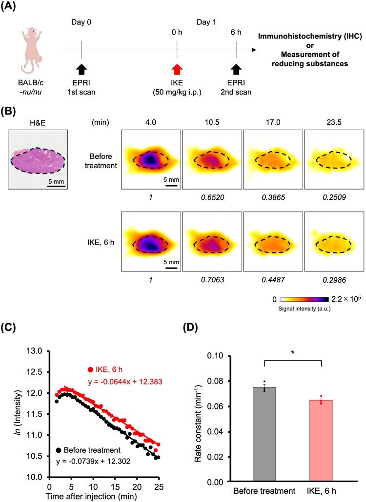
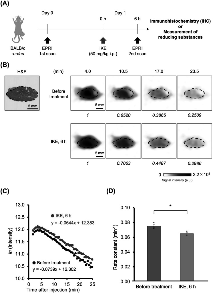
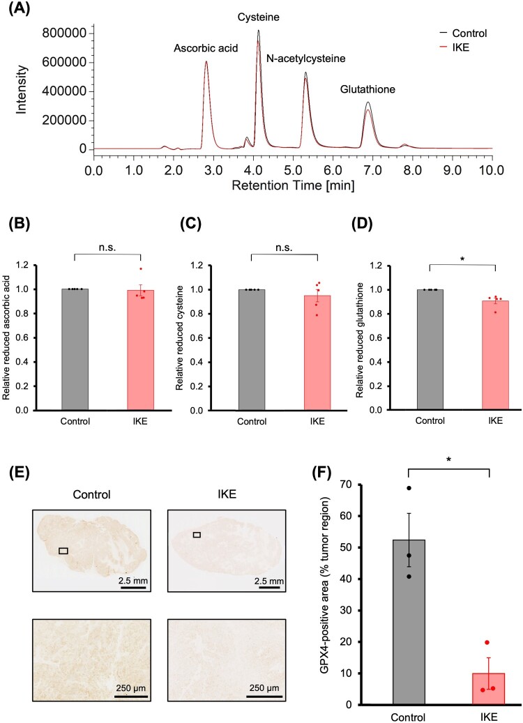
Targeting ferroptosis, cell death caused by the iron-dependent accumulation of lipid peroxides, and disruption of the redox balance are promising strategies in cancer therapy owing to the physiological characteristics of cancer cells. However, the detection of ferroptosis using in vivo imaging remains challenging. We previously reported that redox maps showing the reduction power per unit time of implanted tumor tissues via non-invasive redox imaging using a novel, compact, and portable electron paramagnetic resonance imaging (EPRI) device could be compared with tumor tissue sections. This study aimed to apply the EPRI technique to the in vivo detection of ferroptosis. Notably, redox maps reflecting changes in the redox status of tumors induced by the ferroptosis-inducing agent imidazole ketone erastin (IKE) were compared with the immunohistochemical images of 4-hydroxynonenal (4-HNE) in tumor tissue sections. Our comparison revealed a negative correlation between the reducing power of tumor tissue and the number of 4-HNE-positive cells. Furthermore, the control and IKE-treated groups exhibited significantly different distributions on the correlation map. Therefore, redox imaging using EPRI may contribute to the non-invasive detection of ferroptosis in vivo.
期刊介绍:
Redox Report is a multidisciplinary peer-reviewed open access journal focusing on the role of free radicals, oxidative stress, activated oxygen, perioxidative and redox processes, primarily in the human environment and human pathology. Relevant papers on the animal and plant environment, biology and pathology will also be included.
While emphasis is placed upon methodological and intellectual advances underpinned by new data, the journal offers scope for review, hypotheses, critiques and other forms of discussion.


 分享
分享
 求助内容:
求助内容: 应助结果提醒方式:
 扫码关注我们
