Emma Kearney, David Greenald, Gabriele Matilionyte, Sheila Lane, Melissa D Tharmalingam, Jill Davies, Jan-Bernd Stukenborg, Grace Forsyth, Rod T Mitchell
{"title":"人胎儿和青春期前睾丸组织的生殖细胞定量-当前方法的比较。","authors":"Emma Kearney, David Greenald, Gabriele Matilionyte, Sheila Lane, Melissa D Tharmalingam, Jill Davies, Jan-Bernd Stukenborg, Grace Forsyth, Rod T Mitchell","doi":"10.1530/RAF-24-0116","DOIUrl":null,"url":null,"abstract":"<p><strong>Abstract: </strong>Methods to quantify germ cell number in human immature testicular tissues are essential to evaluate the impact of chemotherapy exposures and to optimise cryopreservation protocols used in fertility preservation for prepubertal boys. Established quantification methods rely on the presence of round tubules within the tissue. However, round tubular cross sections are limited in human prepubertal testicular tissues, especially when using in vitro culture. We aimed to assess whether an alternative method of germ cell quantification would provide similar results to recently established methods, without the requirement for round tubules. Human testicular samples included fetal tissue (exposed in vitro to cisplatin, carboplatin or control) or prepubertal tissue (fresh, cryopreserved, fresh in vitro cultured or cryopreserved in vitro cultured). Immunofluorescence assessed AP2γ (gonocytes) and MAGE-A4 ((pre)spermatogonia) expression. Germ cells were quantified by tubular germ cell density (Method 1), which was compared to methods that require round tubules, including spermatogonial number per round tubular cross section (S/T) (Method 2), fertility index (Method 3) and round tubular germ cell density (Method 4). A correlation analysis between methods was performed. Method 1 is strongly and significantly correlated with Method 2 (r = 0.838, P < 0.0001; r = 0.833, P < 0.0001), Method 3 (r = 0.752, P < 0.001; r = 0.802, P < 0.0001) and Method 4 (r = 0.863, P < 0.0001; r = 0.914, P < 0.0001) for fetal and prepubertal tissues, respectively. Given that Method 1 assess tubules irrespective of shape, it may increase the total number of germ cells available for quantification, validating its use for quantification of human testicular tissue samples where the amount of tissue or presence of round tubules is limited.</p><p><strong>Lay summary: </strong>Chemotherapy can damage cells in the testicles that are required to make sperm, often leading to infertility in males. While options to preserve fertility in adult males are available, there are no established methods for young boys. To investigate how chemotherapy damages these cells and to explore approaches to preserve fertility, we require methods to count the number of cells that can develop into sperm. Existing counting methods involve only counting some of the cells in the tissue, but in young boys, it is often necessary to count all of the cells because the amount of tissue is limited. To overcome this, we counted cells in small pieces of human fetal and prepubertal testicles using an alternative method, which allows all cells to be counted. We found similar results using our method compared to three existing methods, making our method useful for counting cells in fetal and prepubertal testicle samples.</p>","PeriodicalId":101312,"journal":{"name":"Reproduction & fertility","volume":" ","pages":""},"PeriodicalIF":3.4000,"publicationDate":"2025-02-07","publicationTypes":"Journal Article","fieldsOfStudy":null,"isOpenAccess":false,"openAccessPdf":"https://www.ncbi.nlm.nih.gov/pmc/articles/PMC11825158/pdf/","citationCount":"0","resultStr":"{\"title\":\"Germ cell quantification in human fetal and prepubertal testis tissues: a comparison of current methodologies.\",\"authors\":\"Emma Kearney, David Greenald, Gabriele Matilionyte, Sheila Lane, Melissa D Tharmalingam, Jill Davies, Jan-Bernd Stukenborg, Grace Forsyth, Rod T Mitchell\",\"doi\":\"10.1530/RAF-24-0116\",\"DOIUrl\":null,\"url\":null,\"abstract\":\"<p><strong>Abstract: </strong>Methods to quantify germ cell number in human immature testicular tissues are essential to evaluate the impact of chemotherapy exposures and to optimise cryopreservation protocols used in fertility preservation for prepubertal boys. Established quantification methods rely on the presence of round tubules within the tissue. However, round tubular cross sections are limited in human prepubertal testicular tissues, especially when using in vitro culture. We aimed to assess whether an alternative method of germ cell quantification would provide similar results to recently established methods, without the requirement for round tubules. Human testicular samples included fetal tissue (exposed in vitro to cisplatin, carboplatin or control) or prepubertal tissue (fresh, cryopreserved, fresh in vitro cultured or cryopreserved in vitro cultured). Immunofluorescence assessed AP2γ (gonocytes) and MAGE-A4 ((pre)spermatogonia) expression. Germ cells were quantified by tubular germ cell density (Method 1), which was compared to methods that require round tubules, including spermatogonial number per round tubular cross section (S/T) (Method 2), fertility index (Method 3) and round tubular germ cell density (Method 4). A correlation analysis between methods was performed. Method 1 is strongly and significantly correlated with Method 2 (r = 0.838, P < 0.0001; r = 0.833, P < 0.0001), Method 3 (r = 0.752, P < 0.001; r = 0.802, P < 0.0001) and Method 4 (r = 0.863, P < 0.0001; r = 0.914, P < 0.0001) for fetal and prepubertal tissues, respectively. Given that Method 1 assess tubules irrespective of shape, it may increase the total number of germ cells available for quantification, validating its use for quantification of human testicular tissue samples where the amount of tissue or presence of round tubules is limited.</p><p><strong>Lay summary: </strong>Chemotherapy can damage cells in the testicles that are required to make sperm, often leading to infertility in males. While options to preserve fertility in adult males are available, there are no established methods for young boys. To investigate how chemotherapy damages these cells and to explore approaches to preserve fertility, we require methods to count the number of cells that can develop into sperm. Existing counting methods involve only counting some of the cells in the tissue, but in young boys, it is often necessary to count all of the cells because the amount of tissue is limited. To overcome this, we counted cells in small pieces of human fetal and prepubertal testicles using an alternative method, which allows all cells to be counted. We found similar results using our method compared to three existing methods, making our method useful for counting cells in fetal and prepubertal testicle samples.</p>\",\"PeriodicalId\":101312,\"journal\":{\"name\":\"Reproduction & fertility\",\"volume\":\" \",\"pages\":\"\"},\"PeriodicalIF\":3.4000,\"publicationDate\":\"2025-02-07\",\"publicationTypes\":\"Journal Article\",\"fieldsOfStudy\":null,\"isOpenAccess\":false,\"openAccessPdf\":\"https://www.ncbi.nlm.nih.gov/pmc/articles/PMC11825158/pdf/\",\"citationCount\":\"0\",\"resultStr\":null,\"platform\":\"Semanticscholar\",\"paperid\":null,\"PeriodicalName\":\"Reproduction & fertility\",\"FirstCategoryId\":\"1085\",\"ListUrlMain\":\"https://doi.org/10.1530/RAF-24-0116\",\"RegionNum\":0,\"RegionCategory\":null,\"ArticlePicture\":[],\"TitleCN\":null,\"AbstractTextCN\":null,\"PMCID\":null,\"EPubDate\":\"2025/1/1 0:00:00\",\"PubModel\":\"Print\",\"JCR\":\"Q2\",\"JCRName\":\"REPRODUCTIVE BIOLOGY\",\"Score\":null,\"Total\":0}","platform":"Semanticscholar","paperid":null,"PeriodicalName":"Reproduction & fertility","FirstCategoryId":"1085","ListUrlMain":"https://doi.org/10.1530/RAF-24-0116","RegionNum":0,"RegionCategory":null,"ArticlePicture":[],"TitleCN":null,"AbstractTextCN":null,"PMCID":null,"EPubDate":"2025/1/1 0:00:00","PubModel":"Print","JCR":"Q2","JCRName":"REPRODUCTIVE BIOLOGY","Score":null,"Total":0}
引用次数: 0
摘要
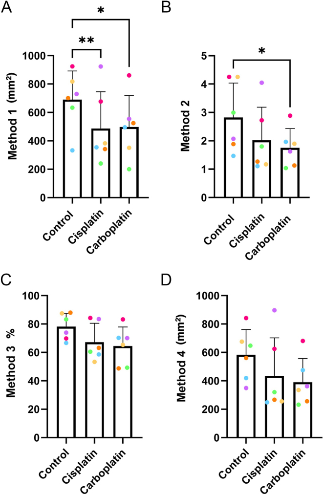
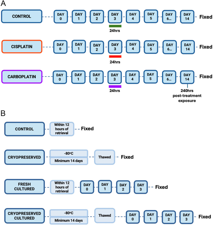
量化人类未成熟睾丸组织中生殖细胞数量的方法对于评估化疗暴露的影响以及优化用于青春期前男孩生育能力保存的冷冻保存方案至关重要。已建立的定量方法依赖于组织内圆管的存在。然而,圆形管状截面在人类青春期前睾丸组织中是有限的,特别是在体外培养时。我们的目的是评估一种生殖细胞定量的替代方法是否会提供与最近建立的方法相似的结果,而不需要圆管。人类睾丸样本包括胎儿组织(在体外暴露于顺铂、卡铂或对照)或青春期前组织(新鲜、冷冻保存、新鲜体外培养或体外冷冻保存)。免疫荧光检测AP2γ(性腺细胞)和MAGE-A4(前)精原细胞)的表达。用管状生殖细胞密度法(方法1)定量生殖细胞,并与需要圆管的方法进行比较,包括:每圆管截面精原细胞数(S/T)(方法2)、生育指数(FI)(方法3)和圆管生殖细胞密度(方法4),并进行方法间的相关性分析。方法1与方法2相关性强且显著(r=0.838, p
Germ cell quantification in human fetal and prepubertal testis tissues: a comparison of current methodologies.
Abstract: Methods to quantify germ cell number in human immature testicular tissues are essential to evaluate the impact of chemotherapy exposures and to optimise cryopreservation protocols used in fertility preservation for prepubertal boys. Established quantification methods rely on the presence of round tubules within the tissue. However, round tubular cross sections are limited in human prepubertal testicular tissues, especially when using in vitro culture. We aimed to assess whether an alternative method of germ cell quantification would provide similar results to recently established methods, without the requirement for round tubules. Human testicular samples included fetal tissue (exposed in vitro to cisplatin, carboplatin or control) or prepubertal tissue (fresh, cryopreserved, fresh in vitro cultured or cryopreserved in vitro cultured). Immunofluorescence assessed AP2γ (gonocytes) and MAGE-A4 ((pre)spermatogonia) expression. Germ cells were quantified by tubular germ cell density (Method 1), which was compared to methods that require round tubules, including spermatogonial number per round tubular cross section (S/T) (Method 2), fertility index (Method 3) and round tubular germ cell density (Method 4). A correlation analysis between methods was performed. Method 1 is strongly and significantly correlated with Method 2 (r = 0.838, P < 0.0001; r = 0.833, P < 0.0001), Method 3 (r = 0.752, P < 0.001; r = 0.802, P < 0.0001) and Method 4 (r = 0.863, P < 0.0001; r = 0.914, P < 0.0001) for fetal and prepubertal tissues, respectively. Given that Method 1 assess tubules irrespective of shape, it may increase the total number of germ cells available for quantification, validating its use for quantification of human testicular tissue samples where the amount of tissue or presence of round tubules is limited.
Lay summary: Chemotherapy can damage cells in the testicles that are required to make sperm, often leading to infertility in males. While options to preserve fertility in adult males are available, there are no established methods for young boys. To investigate how chemotherapy damages these cells and to explore approaches to preserve fertility, we require methods to count the number of cells that can develop into sperm. Existing counting methods involve only counting some of the cells in the tissue, but in young boys, it is often necessary to count all of the cells because the amount of tissue is limited. To overcome this, we counted cells in small pieces of human fetal and prepubertal testicles using an alternative method, which allows all cells to be counted. We found similar results using our method compared to three existing methods, making our method useful for counting cells in fetal and prepubertal testicle samples.


 分享
分享

 求助内容:
求助内容: 应助结果提醒方式:
 扫码关注我们
