{"title":"多平面CT成像对成釉细胞瘤的分类及预后评价:回顾性分析。","authors":"Jo-Eun Kim, Jun-Bum Cho, Won-Jin Yi, Min-Suk Heo, Sam-Sun Lee, Kyung-Hoe Huh","doi":"10.1186/s12903-025-05485-6","DOIUrl":null,"url":null,"abstract":"<p><strong>Background: </strong>Ameloblastoma is the most prevalent odontogenic tumor of the jaw, with a significant recurrence rate. It was conventionally classified radiographically as unilocular or multilocular. As 3D images become more common, there is a need to reassess this classification. This study aimed to conduct a multi-planar assessment of ameloblastoma using CT images, reclassify the lesions based on locularity, and correlate the locularity with imaging characteristics and recurrence.</p><p><strong>Methods: </strong>Panoramic radiographs and CT images of histopathologically confirmed 255 ameloblastoma patients were included. The analysis encompassed imaging features including locularity, expansion, thinning, cortical perforation, and root resorption. Image characteristics associated with recurrence rate were examined, and image features were assessed based on locularity.</p><p><strong>Results: </strong>On panoramic radiographs, most ameloblastomas (68.6%) were classified as multilocular, while 31.4% were unilocular. The CT scan demonstrated that 46.3% of the ameloblastomas were pseudo-multilocular, 29.4% were unilocular, and 24.3% were multilocular. Multilocular cases on panoramic radiographs often appeared as pseudo-multilocular on CT. Cortical perforation, root resorption, and size were significantly correlated with CT locularity types (p < 0.05). The recurrence rate was 14.11%, and the risk of recurrence was substantially reduced by mandibulectomy and a larger tumor size. No other variables were significantly related to recurrence.</p><p><strong>Conclusion: </strong>An accurate diagnosis of ameloblastoma and surgical planning to reduce the recurrence rate are contingent upon the use of three-dimensional imaging modalities, such as CT.</p>","PeriodicalId":9072,"journal":{"name":"BMC Oral Health","volume":"25 1","pages":"115"},"PeriodicalIF":3.1000,"publicationDate":"2025-01-22","publicationTypes":"Journal Article","fieldsOfStudy":null,"isOpenAccess":false,"openAccessPdf":"https://www.ncbi.nlm.nih.gov/pmc/articles/PMC11755854/pdf/","citationCount":"0","resultStr":"{\"title\":\"Classification and prognostic evaluation of ameloblastoma using multiplanar CT imaging: a retrospective analysis.\",\"authors\":\"Jo-Eun Kim, Jun-Bum Cho, Won-Jin Yi, Min-Suk Heo, Sam-Sun Lee, Kyung-Hoe Huh\",\"doi\":\"10.1186/s12903-025-05485-6\",\"DOIUrl\":null,\"url\":null,\"abstract\":\"<p><strong>Background: </strong>Ameloblastoma is the most prevalent odontogenic tumor of the jaw, with a significant recurrence rate. It was conventionally classified radiographically as unilocular or multilocular. As 3D images become more common, there is a need to reassess this classification. This study aimed to conduct a multi-planar assessment of ameloblastoma using CT images, reclassify the lesions based on locularity, and correlate the locularity with imaging characteristics and recurrence.</p><p><strong>Methods: </strong>Panoramic radiographs and CT images of histopathologically confirmed 255 ameloblastoma patients were included. The analysis encompassed imaging features including locularity, expansion, thinning, cortical perforation, and root resorption. Image characteristics associated with recurrence rate were examined, and image features were assessed based on locularity.</p><p><strong>Results: </strong>On panoramic radiographs, most ameloblastomas (68.6%) were classified as multilocular, while 31.4% were unilocular. The CT scan demonstrated that 46.3% of the ameloblastomas were pseudo-multilocular, 29.4% were unilocular, and 24.3% were multilocular. Multilocular cases on panoramic radiographs often appeared as pseudo-multilocular on CT. Cortical perforation, root resorption, and size were significantly correlated with CT locularity types (p < 0.05). The recurrence rate was 14.11%, and the risk of recurrence was substantially reduced by mandibulectomy and a larger tumor size. No other variables were significantly related to recurrence.</p><p><strong>Conclusion: </strong>An accurate diagnosis of ameloblastoma and surgical planning to reduce the recurrence rate are contingent upon the use of three-dimensional imaging modalities, such as CT.</p>\",\"PeriodicalId\":9072,\"journal\":{\"name\":\"BMC Oral Health\",\"volume\":\"25 1\",\"pages\":\"115\"},\"PeriodicalIF\":3.1000,\"publicationDate\":\"2025-01-22\",\"publicationTypes\":\"Journal Article\",\"fieldsOfStudy\":null,\"isOpenAccess\":false,\"openAccessPdf\":\"https://www.ncbi.nlm.nih.gov/pmc/articles/PMC11755854/pdf/\",\"citationCount\":\"0\",\"resultStr\":null,\"platform\":\"Semanticscholar\",\"paperid\":null,\"PeriodicalName\":\"BMC Oral Health\",\"FirstCategoryId\":\"3\",\"ListUrlMain\":\"https://doi.org/10.1186/s12903-025-05485-6\",\"RegionNum\":2,\"RegionCategory\":\"医学\",\"ArticlePicture\":[],\"TitleCN\":null,\"AbstractTextCN\":null,\"PMCID\":null,\"EPubDate\":\"\",\"PubModel\":\"\",\"JCR\":\"Q1\",\"JCRName\":\"DENTISTRY, ORAL SURGERY & MEDICINE\",\"Score\":null,\"Total\":0}","platform":"Semanticscholar","paperid":null,"PeriodicalName":"BMC Oral Health","FirstCategoryId":"3","ListUrlMain":"https://doi.org/10.1186/s12903-025-05485-6","RegionNum":2,"RegionCategory":"医学","ArticlePicture":[],"TitleCN":null,"AbstractTextCN":null,"PMCID":null,"EPubDate":"","PubModel":"","JCR":"Q1","JCRName":"DENTISTRY, ORAL SURGERY & MEDICINE","Score":null,"Total":0}
Classification and prognostic evaluation of ameloblastoma using multiplanar CT imaging: a retrospective analysis.
Background: Ameloblastoma is the most prevalent odontogenic tumor of the jaw, with a significant recurrence rate. It was conventionally classified radiographically as unilocular or multilocular. As 3D images become more common, there is a need to reassess this classification. This study aimed to conduct a multi-planar assessment of ameloblastoma using CT images, reclassify the lesions based on locularity, and correlate the locularity with imaging characteristics and recurrence.
Methods: Panoramic radiographs and CT images of histopathologically confirmed 255 ameloblastoma patients were included. The analysis encompassed imaging features including locularity, expansion, thinning, cortical perforation, and root resorption. Image characteristics associated with recurrence rate were examined, and image features were assessed based on locularity.
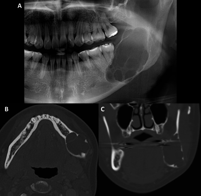
Results: On panoramic radiographs, most ameloblastomas (68.6%) were classified as multilocular, while 31.4% were unilocular. The CT scan demonstrated that 46.3% of the ameloblastomas were pseudo-multilocular, 29.4% were unilocular, and 24.3% were multilocular. Multilocular cases on panoramic radiographs often appeared as pseudo-multilocular on CT. Cortical perforation, root resorption, and size were significantly correlated with CT locularity types (p < 0.05). The recurrence rate was 14.11%, and the risk of recurrence was substantially reduced by mandibulectomy and a larger tumor size. No other variables were significantly related to recurrence.
Conclusion: An accurate diagnosis of ameloblastoma and surgical planning to reduce the recurrence rate are contingent upon the use of three-dimensional imaging modalities, such as CT.
期刊介绍:
BMC Oral Health is an open access, peer-reviewed journal that considers articles on all aspects of the prevention, diagnosis and management of disorders of the mouth, teeth and gums, as well as related molecular genetics, pathophysiology, and epidemiology.







 京公网安备 11010802042870号
京ICP备2023020795号-1
京公网安备 11010802042870号
京ICP备2023020795号-1

 分享
分享
 求助内容:
求助内容: 应助结果提醒方式:
 扫码关注我们
