{"title":"硬膜内腰椎间盘突出症术后炎症所致脊髓空洞伴中央管扩大。","authors":"Hiroki Hagizawa, Yu Moriguchi, Tomonori Yamada, Masafumi Kashii","doi":"10.2176/jns-nmc.2024-0165","DOIUrl":null,"url":null,"abstract":"<p><p>Intradural lumbar disc herniation (ILDH) is a very rare condition, with cerebrospinal fluid (CSF) leakage as a postoperative complication. The central canal of the conus medullaris was reported to communicate with the subarachnoid space through a caudal aperture; however, this aperture has never been observed in vivo. Herein, we report a case of L1/2 ILDH with postoperative spinal adhesive arachnoiditis and syringomyelia in which the communication considered to be a caudal aperture was detected. A 67-year-old woman complained of acute lower back pain and right leg pain with muscle weakness. Magnetic resonance imaging (MRI) revealed a large mass in the spinal canal at the L1/2 level. Intraoperatively, no herniated fragments were found in the epidural space. Thus, ILDH was suspected, and an intradural examination revealed tightly adherent herniated fragments in the swollen cauda equina and surrounding neovascularization. Postoperatively, symptoms improved once; however, 3 months later, severe bilateral leg muscle weakness and urinary retention developed. She was diagnosed with postoperative syringomyelia by MRI, and myelography and follow-up computed tomography was performed. Immediately after the administration of nonionic contrast agents, the syrinx of conus and epiconus were delineated through the central canal, and the communication between the central canal and the subarachnoid space could be visualized. Symptoms improved after syringo-subarachnoid shunt and duroplasty. It is likely that when CSF perfusion was impaired because of adhesive arachnoiditis around the conus medullaris, the central canal, which had been obstructed with growth, reopened, and the patient presented with syringomyelia.</p>","PeriodicalId":101331,"journal":{"name":"NMC case report journal","volume":"11 ","pages":"421-425"},"PeriodicalIF":0.0000,"publicationDate":"2024-12-25","publicationTypes":"Journal Article","fieldsOfStudy":null,"isOpenAccess":false,"openAccessPdf":"https://www.ncbi.nlm.nih.gov/pmc/articles/PMC11756898/pdf/","citationCount":"0","resultStr":"{\"title\":\"Syringomyelia with Central Canal Enlargement Caused by Postoperative Inflammation after Surgery for Intradural Lumbar Disc Herniation.\",\"authors\":\"Hiroki Hagizawa, Yu Moriguchi, Tomonori Yamada, Masafumi Kashii\",\"doi\":\"10.2176/jns-nmc.2024-0165\",\"DOIUrl\":null,\"url\":null,\"abstract\":\"<p><p>Intradural lumbar disc herniation (ILDH) is a very rare condition, with cerebrospinal fluid (CSF) leakage as a postoperative complication. The central canal of the conus medullaris was reported to communicate with the subarachnoid space through a caudal aperture; however, this aperture has never been observed in vivo. Herein, we report a case of L1/2 ILDH with postoperative spinal adhesive arachnoiditis and syringomyelia in which the communication considered to be a caudal aperture was detected. A 67-year-old woman complained of acute lower back pain and right leg pain with muscle weakness. Magnetic resonance imaging (MRI) revealed a large mass in the spinal canal at the L1/2 level. Intraoperatively, no herniated fragments were found in the epidural space. Thus, ILDH was suspected, and an intradural examination revealed tightly adherent herniated fragments in the swollen cauda equina and surrounding neovascularization. Postoperatively, symptoms improved once; however, 3 months later, severe bilateral leg muscle weakness and urinary retention developed. She was diagnosed with postoperative syringomyelia by MRI, and myelography and follow-up computed tomography was performed. Immediately after the administration of nonionic contrast agents, the syrinx of conus and epiconus were delineated through the central canal, and the communication between the central canal and the subarachnoid space could be visualized. Symptoms improved after syringo-subarachnoid shunt and duroplasty. It is likely that when CSF perfusion was impaired because of adhesive arachnoiditis around the conus medullaris, the central canal, which had been obstructed with growth, reopened, and the patient presented with syringomyelia.</p>\",\"PeriodicalId\":101331,\"journal\":{\"name\":\"NMC case report journal\",\"volume\":\"11 \",\"pages\":\"421-425\"},\"PeriodicalIF\":0.0000,\"publicationDate\":\"2024-12-25\",\"publicationTypes\":\"Journal Article\",\"fieldsOfStudy\":null,\"isOpenAccess\":false,\"openAccessPdf\":\"https://www.ncbi.nlm.nih.gov/pmc/articles/PMC11756898/pdf/\",\"citationCount\":\"0\",\"resultStr\":null,\"platform\":\"Semanticscholar\",\"paperid\":null,\"PeriodicalName\":\"NMC case report journal\",\"FirstCategoryId\":\"1085\",\"ListUrlMain\":\"https://doi.org/10.2176/jns-nmc.2024-0165\",\"RegionNum\":0,\"RegionCategory\":null,\"ArticlePicture\":[],\"TitleCN\":null,\"AbstractTextCN\":null,\"PMCID\":null,\"EPubDate\":\"2024/1/1 0:00:00\",\"PubModel\":\"eCollection\",\"JCR\":\"\",\"JCRName\":\"\",\"Score\":null,\"Total\":0}","platform":"Semanticscholar","paperid":null,"PeriodicalName":"NMC case report journal","FirstCategoryId":"1085","ListUrlMain":"https://doi.org/10.2176/jns-nmc.2024-0165","RegionNum":0,"RegionCategory":null,"ArticlePicture":[],"TitleCN":null,"AbstractTextCN":null,"PMCID":null,"EPubDate":"2024/1/1 0:00:00","PubModel":"eCollection","JCR":"","JCRName":"","Score":null,"Total":0}
Syringomyelia with Central Canal Enlargement Caused by Postoperative Inflammation after Surgery for Intradural Lumbar Disc Herniation.
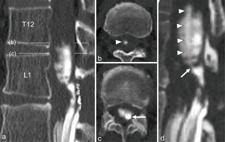
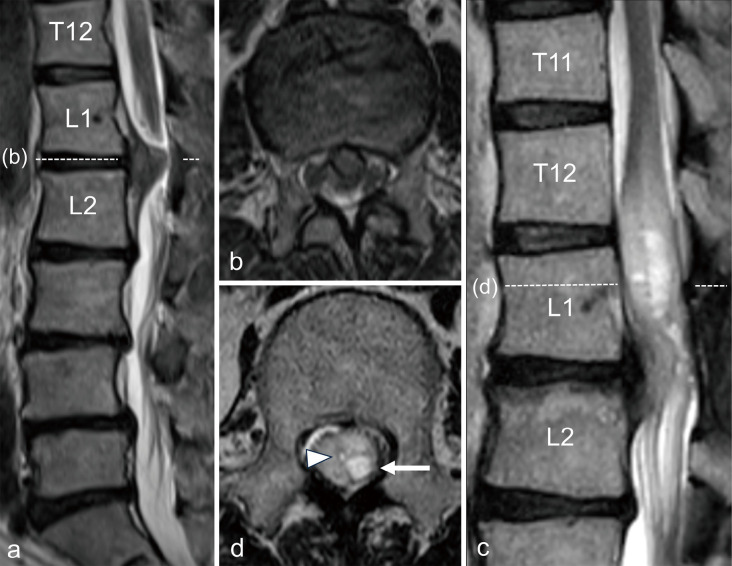
Intradural lumbar disc herniation (ILDH) is a very rare condition, with cerebrospinal fluid (CSF) leakage as a postoperative complication. The central canal of the conus medullaris was reported to communicate with the subarachnoid space through a caudal aperture; however, this aperture has never been observed in vivo. Herein, we report a case of L1/2 ILDH with postoperative spinal adhesive arachnoiditis and syringomyelia in which the communication considered to be a caudal aperture was detected. A 67-year-old woman complained of acute lower back pain and right leg pain with muscle weakness. Magnetic resonance imaging (MRI) revealed a large mass in the spinal canal at the L1/2 level. Intraoperatively, no herniated fragments were found in the epidural space. Thus, ILDH was suspected, and an intradural examination revealed tightly adherent herniated fragments in the swollen cauda equina and surrounding neovascularization. Postoperatively, symptoms improved once; however, 3 months later, severe bilateral leg muscle weakness and urinary retention developed. She was diagnosed with postoperative syringomyelia by MRI, and myelography and follow-up computed tomography was performed. Immediately after the administration of nonionic contrast agents, the syrinx of conus and epiconus were delineated through the central canal, and the communication between the central canal and the subarachnoid space could be visualized. Symptoms improved after syringo-subarachnoid shunt and duroplasty. It is likely that when CSF perfusion was impaired because of adhesive arachnoiditis around the conus medullaris, the central canal, which had been obstructed with growth, reopened, and the patient presented with syringomyelia.





 京公网安备 11010802042870号
京ICP备2023020795号-1
京公网安备 11010802042870号
京ICP备2023020795号-1

 分享
分享

 求助内容:
求助内容: 应助结果提醒方式:
 扫码关注我们
