{"title":"外侧半月板前交叉韧带异常插入1例。","authors":"Mackenzie Norman, Katie Zehner, Elizabeth Gardner","doi":"10.2106/JBJS.CC.24.00395","DOIUrl":null,"url":null,"abstract":"<p><strong>Case: </strong>A 16-year-old woman presented with acute on chronic knee pain and instability following a twisting injury. The tibial insertion of the anterior cruciate ligament (ACL) was nonvisualized on magnetic resonance imaging. A cord-like ACL, originating from the lateral intercondylar notch and inserting smoothly into the anterior horn of the intact lateral meniscus, was found on arthroscopy. Her ACL was reconstructed with quadriceps tendon autograft.</p><p><strong>Conclusion: </strong>We report a rare case of dysplastic ACL inserting into the lateral meniscus in a patient without obvious skeletal deformities. This pathology could be congenital or traumatic. Surgeons and radiologists should be aware of this rare variant.</p>","PeriodicalId":14748,"journal":{"name":"JBJS case connector","volume":"15 1","pages":""},"PeriodicalIF":0.0000,"publicationDate":"2025-01-24","publicationTypes":"Journal Article","fieldsOfStudy":null,"isOpenAccess":false,"openAccessPdf":"https://www.ncbi.nlm.nih.gov/pmc/articles/PMC11756712/pdf/","citationCount":"0","resultStr":"{\"title\":\"Aberrant Insertion of the Anterior Cruciate Ligament on the Lateral Meniscus: A Case Report.\",\"authors\":\"Mackenzie Norman, Katie Zehner, Elizabeth Gardner\",\"doi\":\"10.2106/JBJS.CC.24.00395\",\"DOIUrl\":null,\"url\":null,\"abstract\":\"<p><strong>Case: </strong>A 16-year-old woman presented with acute on chronic knee pain and instability following a twisting injury. The tibial insertion of the anterior cruciate ligament (ACL) was nonvisualized on magnetic resonance imaging. A cord-like ACL, originating from the lateral intercondylar notch and inserting smoothly into the anterior horn of the intact lateral meniscus, was found on arthroscopy. Her ACL was reconstructed with quadriceps tendon autograft.</p><p><strong>Conclusion: </strong>We report a rare case of dysplastic ACL inserting into the lateral meniscus in a patient without obvious skeletal deformities. This pathology could be congenital or traumatic. Surgeons and radiologists should be aware of this rare variant.</p>\",\"PeriodicalId\":14748,\"journal\":{\"name\":\"JBJS case connector\",\"volume\":\"15 1\",\"pages\":\"\"},\"PeriodicalIF\":0.0000,\"publicationDate\":\"2025-01-24\",\"publicationTypes\":\"Journal Article\",\"fieldsOfStudy\":null,\"isOpenAccess\":false,\"openAccessPdf\":\"https://www.ncbi.nlm.nih.gov/pmc/articles/PMC11756712/pdf/\",\"citationCount\":\"0\",\"resultStr\":null,\"platform\":\"Semanticscholar\",\"paperid\":null,\"PeriodicalName\":\"JBJS case connector\",\"FirstCategoryId\":\"1085\",\"ListUrlMain\":\"https://doi.org/10.2106/JBJS.CC.24.00395\",\"RegionNum\":0,\"RegionCategory\":null,\"ArticlePicture\":[],\"TitleCN\":null,\"AbstractTextCN\":null,\"PMCID\":null,\"EPubDate\":\"2025/1/1 0:00:00\",\"PubModel\":\"eCollection\",\"JCR\":\"Q4\",\"JCRName\":\"Medicine\",\"Score\":null,\"Total\":0}","platform":"Semanticscholar","paperid":null,"PeriodicalName":"JBJS case connector","FirstCategoryId":"1085","ListUrlMain":"https://doi.org/10.2106/JBJS.CC.24.00395","RegionNum":0,"RegionCategory":null,"ArticlePicture":[],"TitleCN":null,"AbstractTextCN":null,"PMCID":null,"EPubDate":"2025/1/1 0:00:00","PubModel":"eCollection","JCR":"Q4","JCRName":"Medicine","Score":null,"Total":0}
Aberrant Insertion of the Anterior Cruciate Ligament on the Lateral Meniscus: A Case Report.
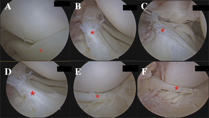
Case: A 16-year-old woman presented with acute on chronic knee pain and instability following a twisting injury. The tibial insertion of the anterior cruciate ligament (ACL) was nonvisualized on magnetic resonance imaging. A cord-like ACL, originating from the lateral intercondylar notch and inserting smoothly into the anterior horn of the intact lateral meniscus, was found on arthroscopy. Her ACL was reconstructed with quadriceps tendon autograft.
Conclusion: We report a rare case of dysplastic ACL inserting into the lateral meniscus in a patient without obvious skeletal deformities. This pathology could be congenital or traumatic. Surgeons and radiologists should be aware of this rare variant.
期刊介绍:
JBJS Case Connector helps improve patient care by providing the medical community with a journal that harnesses technology to provide information tools for discovery and reporting of unusual musculoskeletal problems, findings, treatment, and outcomes. Co-edited by Thomas W. Bauer, MD, PhD, and Ronald W. Lindsey, MD, JBJS Case Connector assists orthopaedic surgeons in the search for precedents, connections, and trends in their efforts to improve patient care. Using this unique journal, surgeons can find the commonalities between cases, benefit from the experience of their peers, and filter case information by many important variables in order to provide the best possible care for orthopaedic patients. This cross-referenced online journal includes thousands of orthopaedic case reports. It compiles symptoms, conditions, and demographic details to empower surgeons to find cases similar to theirs. Surgeons can mine the database to reveal emerging trends and identify patterns, distinguishing between truly rare cases and repeated, related single instances of a larger problem. The JBJS Case Connector Image Quiz feature provides interactive quizzes based on images from content published by JBJS and includes a discussion area for further exploration of ideas and concepts. The JBJS Image Quiz app for iPad and iPhone is available in the App Store. Contributions to JBJS Case Connector are welcomed from anywhere in the world and are considered on their merits. Articles must be written in English and should be submitted as outlined in the Instructions to Authors. All authors must abide by the JBJS ethics policies and all submissions to JBJS Case Connector are covered by the JBJS embargo policy.







 京公网安备 11010802042870号
京ICP备2023020795号-1
京公网安备 11010802042870号
京ICP备2023020795号-1

 分享
分享
 求助内容:
求助内容: 应助结果提醒方式:
 扫码关注我们
