{"title":"评估支气管肺泡灌洗液和外周血淋巴细胞亚群对各种弥漫性间质性肺病亚型的诊断价值。","authors":"Sonoko Harada, Motoyasu Kato, Kazuyuki Nakagome, Hitoshi Sasano, Yuki Tanabe, Tomohito Takeshige, Yuuki Sandhu, Kei Matsuno, Shoko Ueda, Sumiko Abe, Takayasu Nishimaki, Shun Shinomiya, Jun Ito, Sachiko Miyake, Ko Okumura, Makoto Nagata, Kazuhisa Takahashi, Norihiro Harada","doi":"10.3390/biom15010122","DOIUrl":null,"url":null,"abstract":"<p><p>Diffuse interstitial lung diseases (ILD) include conditions with identifiable causes such as chronic eosinophilic pneumonia (CEP), sarcoidosis (SAR), chronic hypersensitivity pneumonitis (CHP), and connective tissue disease-associated interstitial pneumonia (CTD), as well as idiopathic interstitial pneumonia (IIP) of unknown origin. In non-IIP diffuse lung diseases, bronchoalveolar lavage (BAL) fluid appearance is diagnostic. This study examines lymphocyte subsets in BAL fluid and peripheral blood of 56 patients with diffuse ILD, excluding idiopathic pulmonary fibrosis (IPF), who underwent BAL for diagnostic purposes. Patients were classified into CEP, SAR, CHP, CTD, and IIP groups, and clinical data, BAL cell analysis, and peripheral blood mononuclear cell analysis were compared. Eosinophils and type 3 innate lymphocytes (ILC3s) were significantly increased in the BAL fluid of the CEP group. Receiver operating characteristic curve analysis identified eosinophils ≥ 8% in BAL cells and ILC3s ≥ 0.0176% in the BAL lymphocyte fraction as thresholds distinguishing CEP. SAR patients exhibited significantly elevated CD4/CD8 ratios in the BAL fluid, with a ratio of 3.95 or higher and type 1 innate lymphoid cell frequency ≥ 0.254% as differentiation markers. High Th1 cell frequency (≥17.4%) in BAL lymphocytes in IIP, elevated serum KL-6 (≥2081 U/mL) and SP-D (≥261 ng/mL) in CHP, and increased BAL neutrophils (≥2.0%) or a low CD4/CD8 ratio (≤1.2) in CTD serve as distinguishing markers for each ILD. Excluding CEP and SAR, CD4<sup>+</sup> T cell frequencies, including Th1, Th17, and Treg cells in peripheral blood, may differentiate IIP, CHP, and CTD.</p>","PeriodicalId":8943,"journal":{"name":"Biomolecules","volume":"15 1","pages":""},"PeriodicalIF":4.8000,"publicationDate":"2025-01-14","publicationTypes":"Journal Article","fieldsOfStudy":null,"isOpenAccess":false,"openAccessPdf":"https://www.ncbi.nlm.nih.gov/pmc/articles/PMC11763757/pdf/","citationCount":"0","resultStr":"{\"title\":\"Evaluating the Diagnostic Value of Lymphocyte Subsets in Bronchoalveolar Lavage Fluid and Peripheral Blood Across Various Diffuse Interstitial Lung Disease Subtypes.\",\"authors\":\"Sonoko Harada, Motoyasu Kato, Kazuyuki Nakagome, Hitoshi Sasano, Yuki Tanabe, Tomohito Takeshige, Yuuki Sandhu, Kei Matsuno, Shoko Ueda, Sumiko Abe, Takayasu Nishimaki, Shun Shinomiya, Jun Ito, Sachiko Miyake, Ko Okumura, Makoto Nagata, Kazuhisa Takahashi, Norihiro Harada\",\"doi\":\"10.3390/biom15010122\",\"DOIUrl\":null,\"url\":null,\"abstract\":\"<p><p>Diffuse interstitial lung diseases (ILD) include conditions with identifiable causes such as chronic eosinophilic pneumonia (CEP), sarcoidosis (SAR), chronic hypersensitivity pneumonitis (CHP), and connective tissue disease-associated interstitial pneumonia (CTD), as well as idiopathic interstitial pneumonia (IIP) of unknown origin. In non-IIP diffuse lung diseases, bronchoalveolar lavage (BAL) fluid appearance is diagnostic. This study examines lymphocyte subsets in BAL fluid and peripheral blood of 56 patients with diffuse ILD, excluding idiopathic pulmonary fibrosis (IPF), who underwent BAL for diagnostic purposes. Patients were classified into CEP, SAR, CHP, CTD, and IIP groups, and clinical data, BAL cell analysis, and peripheral blood mononuclear cell analysis were compared. Eosinophils and type 3 innate lymphocytes (ILC3s) were significantly increased in the BAL fluid of the CEP group. Receiver operating characteristic curve analysis identified eosinophils ≥ 8% in BAL cells and ILC3s ≥ 0.0176% in the BAL lymphocyte fraction as thresholds distinguishing CEP. SAR patients exhibited significantly elevated CD4/CD8 ratios in the BAL fluid, with a ratio of 3.95 or higher and type 1 innate lymphoid cell frequency ≥ 0.254% as differentiation markers. High Th1 cell frequency (≥17.4%) in BAL lymphocytes in IIP, elevated serum KL-6 (≥2081 U/mL) and SP-D (≥261 ng/mL) in CHP, and increased BAL neutrophils (≥2.0%) or a low CD4/CD8 ratio (≤1.2) in CTD serve as distinguishing markers for each ILD. Excluding CEP and SAR, CD4<sup>+</sup> T cell frequencies, including Th1, Th17, and Treg cells in peripheral blood, may differentiate IIP, CHP, and CTD.</p>\",\"PeriodicalId\":8943,\"journal\":{\"name\":\"Biomolecules\",\"volume\":\"15 1\",\"pages\":\"\"},\"PeriodicalIF\":4.8000,\"publicationDate\":\"2025-01-14\",\"publicationTypes\":\"Journal Article\",\"fieldsOfStudy\":null,\"isOpenAccess\":false,\"openAccessPdf\":\"https://www.ncbi.nlm.nih.gov/pmc/articles/PMC11763757/pdf/\",\"citationCount\":\"0\",\"resultStr\":null,\"platform\":\"Semanticscholar\",\"paperid\":null,\"PeriodicalName\":\"Biomolecules\",\"FirstCategoryId\":\"99\",\"ListUrlMain\":\"https://doi.org/10.3390/biom15010122\",\"RegionNum\":2,\"RegionCategory\":\"生物学\",\"ArticlePicture\":[],\"TitleCN\":null,\"AbstractTextCN\":null,\"PMCID\":null,\"EPubDate\":\"\",\"PubModel\":\"\",\"JCR\":\"Q1\",\"JCRName\":\"BIOCHEMISTRY & MOLECULAR BIOLOGY\",\"Score\":null,\"Total\":0}","platform":"Semanticscholar","paperid":null,"PeriodicalName":"Biomolecules","FirstCategoryId":"99","ListUrlMain":"https://doi.org/10.3390/biom15010122","RegionNum":2,"RegionCategory":"生物学","ArticlePicture":[],"TitleCN":null,"AbstractTextCN":null,"PMCID":null,"EPubDate":"","PubModel":"","JCR":"Q1","JCRName":"BIOCHEMISTRY & MOLECULAR BIOLOGY","Score":null,"Total":0}
Evaluating the Diagnostic Value of Lymphocyte Subsets in Bronchoalveolar Lavage Fluid and Peripheral Blood Across Various Diffuse Interstitial Lung Disease Subtypes.
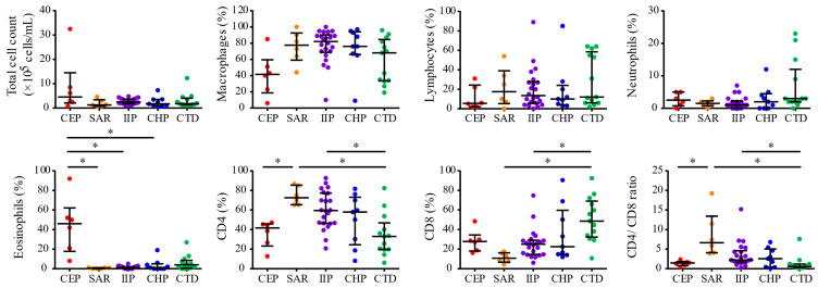
Diffuse interstitial lung diseases (ILD) include conditions with identifiable causes such as chronic eosinophilic pneumonia (CEP), sarcoidosis (SAR), chronic hypersensitivity pneumonitis (CHP), and connective tissue disease-associated interstitial pneumonia (CTD), as well as idiopathic interstitial pneumonia (IIP) of unknown origin. In non-IIP diffuse lung diseases, bronchoalveolar lavage (BAL) fluid appearance is diagnostic. This study examines lymphocyte subsets in BAL fluid and peripheral blood of 56 patients with diffuse ILD, excluding idiopathic pulmonary fibrosis (IPF), who underwent BAL for diagnostic purposes. Patients were classified into CEP, SAR, CHP, CTD, and IIP groups, and clinical data, BAL cell analysis, and peripheral blood mononuclear cell analysis were compared. Eosinophils and type 3 innate lymphocytes (ILC3s) were significantly increased in the BAL fluid of the CEP group. Receiver operating characteristic curve analysis identified eosinophils ≥ 8% in BAL cells and ILC3s ≥ 0.0176% in the BAL lymphocyte fraction as thresholds distinguishing CEP. SAR patients exhibited significantly elevated CD4/CD8 ratios in the BAL fluid, with a ratio of 3.95 or higher and type 1 innate lymphoid cell frequency ≥ 0.254% as differentiation markers. High Th1 cell frequency (≥17.4%) in BAL lymphocytes in IIP, elevated serum KL-6 (≥2081 U/mL) and SP-D (≥261 ng/mL) in CHP, and increased BAL neutrophils (≥2.0%) or a low CD4/CD8 ratio (≤1.2) in CTD serve as distinguishing markers for each ILD. Excluding CEP and SAR, CD4+ T cell frequencies, including Th1, Th17, and Treg cells in peripheral blood, may differentiate IIP, CHP, and CTD.
BiomoleculesBiochemistry, Genetics and Molecular Biology-Molecular Biology
CiteScore
9.40
自引率
3.60%
发文量
1640
审稿时长
18.28 days
期刊介绍:
Biomolecules (ISSN 2218-273X) is an international, peer-reviewed open access journal focusing on biogenic substances and their biological functions, structures, interactions with other molecules, and their microenvironment as well as biological systems. Biomolecules publishes reviews, regular research papers and short communications. Our aim is to encourage scientists to publish their experimental and theoretical results in as much detail as possible. There is no restriction on the length of the papers. The full experimental details must be provided so that the results can be reproduced.






 京公网安备 11010802042870号
京ICP备2023020795号-1
京公网安备 11010802042870号
京ICP备2023020795号-1

 分享
分享
 求助内容:
求助内容: 应助结果提醒方式:
 扫码关注我们
