Lígia Pereira da Silva, Alicia López-Solache, Urbano Santana-Penín, José López-Cedrún, María Jesus Mora, Pablo Varela-Centelles, Antonio González-Mosquera, Almudena Rodríguez-Fernández, Urbano Santana Mora
{"title":"临床牙中线移位不能预测下颌偏短:锥束计算机断层诊断研究。","authors":"Lígia Pereira da Silva, Alicia López-Solache, Urbano Santana-Penín, José López-Cedrún, María Jesus Mora, Pablo Varela-Centelles, Antonio González-Mosquera, Almudena Rodríguez-Fernández, Urbano Santana Mora","doi":"10.3390/diagnostics15020161","DOIUrl":null,"url":null,"abstract":"<p><p><b>Background/Objectives</b>: Interincisive midline deviation is frequent. Determining the cause (dental versus skeletal) is crucial for treatment planning. This study assessed the null hypothesis that neither clinical dental midline shift nor the temporomandibular disorder (TMD)-affected side correlate with maxillary/mandibular asymmetry. <b>Methods</b>: Thirty-eight CBCT scans were analyzed: thirty-five (92.1%) females, three (7.9%) males; mean (SD) age 34.6 (11.9) years old. Tomographic images were acquired using the i-CAT<sup>®</sup> Imaging System; mandibular/maxillary measurements were obtained with the Planmeca Romexis<sup>®</sup> software v.6.This is an ancillary study of a clinical trial (NCT02144233) that included chronic pain (TMD diagnosis; DC/TMD criteria), fully dentate, and stable normo-occlusion participants. <b>Results</b>: We found sixteen (42.1%) dental midline deviations to the right and thirteen (34.2%) to the left. In the study population, the right side was more developed: a hemimandible length of 119.4 (5.7) mm versus 118.6 (5.3) mm for the right and left sides, respectively (95% CI 0.21 to 1.51), <i>p</i> = 0.01. <b>Conclusions</b>: Neither the dental midline shift side nor the affected side predicted a less developed hemimandible.</p>","PeriodicalId":11225,"journal":{"name":"Diagnostics","volume":"15 2","pages":""},"PeriodicalIF":3.8000,"publicationDate":"2025-01-13","publicationTypes":"Journal Article","fieldsOfStudy":null,"isOpenAccess":false,"openAccessPdf":"https://www.ncbi.nlm.nih.gov/pmc/articles/PMC11763747/pdf/","citationCount":"0","resultStr":"{\"title\":\"Clinical Dental Midline Shift Is Not a Predictor of the Side of Shorter Hemimandible: A Cone Beam Computed Tomography Diagnostic Study.\",\"authors\":\"Lígia Pereira da Silva, Alicia López-Solache, Urbano Santana-Penín, José López-Cedrún, María Jesus Mora, Pablo Varela-Centelles, Antonio González-Mosquera, Almudena Rodríguez-Fernández, Urbano Santana Mora\",\"doi\":\"10.3390/diagnostics15020161\",\"DOIUrl\":null,\"url\":null,\"abstract\":\"<p><p><b>Background/Objectives</b>: Interincisive midline deviation is frequent. Determining the cause (dental versus skeletal) is crucial for treatment planning. This study assessed the null hypothesis that neither clinical dental midline shift nor the temporomandibular disorder (TMD)-affected side correlate with maxillary/mandibular asymmetry. <b>Methods</b>: Thirty-eight CBCT scans were analyzed: thirty-five (92.1%) females, three (7.9%) males; mean (SD) age 34.6 (11.9) years old. Tomographic images were acquired using the i-CAT<sup>®</sup> Imaging System; mandibular/maxillary measurements were obtained with the Planmeca Romexis<sup>®</sup> software v.6.This is an ancillary study of a clinical trial (NCT02144233) that included chronic pain (TMD diagnosis; DC/TMD criteria), fully dentate, and stable normo-occlusion participants. <b>Results</b>: We found sixteen (42.1%) dental midline deviations to the right and thirteen (34.2%) to the left. In the study population, the right side was more developed: a hemimandible length of 119.4 (5.7) mm versus 118.6 (5.3) mm for the right and left sides, respectively (95% CI 0.21 to 1.51), <i>p</i> = 0.01. <b>Conclusions</b>: Neither the dental midline shift side nor the affected side predicted a less developed hemimandible.</p>\",\"PeriodicalId\":11225,\"journal\":{\"name\":\"Diagnostics\",\"volume\":\"15 2\",\"pages\":\"\"},\"PeriodicalIF\":3.8000,\"publicationDate\":\"2025-01-13\",\"publicationTypes\":\"Journal Article\",\"fieldsOfStudy\":null,\"isOpenAccess\":false,\"openAccessPdf\":\"https://www.ncbi.nlm.nih.gov/pmc/articles/PMC11763747/pdf/\",\"citationCount\":\"0\",\"resultStr\":null,\"platform\":\"Semanticscholar\",\"paperid\":null,\"PeriodicalName\":\"Diagnostics\",\"FirstCategoryId\":\"3\",\"ListUrlMain\":\"https://doi.org/10.3390/diagnostics15020161\",\"RegionNum\":3,\"RegionCategory\":\"医学\",\"ArticlePicture\":[],\"TitleCN\":null,\"AbstractTextCN\":null,\"PMCID\":null,\"EPubDate\":\"\",\"PubModel\":\"\",\"JCR\":\"Q1\",\"JCRName\":\"MEDICINE, GENERAL & INTERNAL\",\"Score\":null,\"Total\":0}","platform":"Semanticscholar","paperid":null,"PeriodicalName":"Diagnostics","FirstCategoryId":"3","ListUrlMain":"https://doi.org/10.3390/diagnostics15020161","RegionNum":3,"RegionCategory":"医学","ArticlePicture":[],"TitleCN":null,"AbstractTextCN":null,"PMCID":null,"EPubDate":"","PubModel":"","JCR":"Q1","JCRName":"MEDICINE, GENERAL & INTERNAL","Score":null,"Total":0}
引用次数: 0
摘要
背景/目的:切点间中线偏移频繁。确定病因(牙齿还是骨骼)对治疗计划至关重要。本研究评估了临床牙中线移位和颞下颌紊乱(TMD)影响侧与上颌/下颌不对称无关的零假设。方法:分析38例CBCT扫描:女性35例(92.1%),男性3例(7.9%);平均(SD)年龄为34.6(11.9)岁。使用i-CAT®成像系统获取层析图像;下颌/上颌测量使用Planmeca Romexis®软件v.6进行。这是一项临床试验(NCT02144233)的辅助研究,其中包括慢性疼痛(TMD诊断;DC/TMD标准),全齿,稳定的正常咬合参与者。结果:牙中线偏右16例(42.1%),偏左13例(34.2%)。在研究人群中,右侧更为发达:右侧和左侧的下颌骨长度分别为119.4 (5.7)mm和118.6 (5.3)mm (95% CI 0.21至1.51),p = 0.01。结论:牙中线移位侧和患侧均不能预测下颌骨发育不全。
Clinical Dental Midline Shift Is Not a Predictor of the Side of Shorter Hemimandible: A Cone Beam Computed Tomography Diagnostic Study.
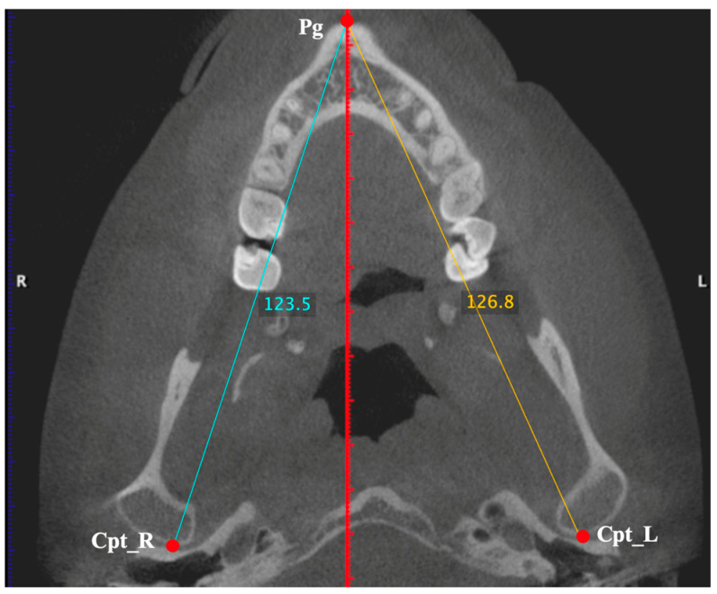
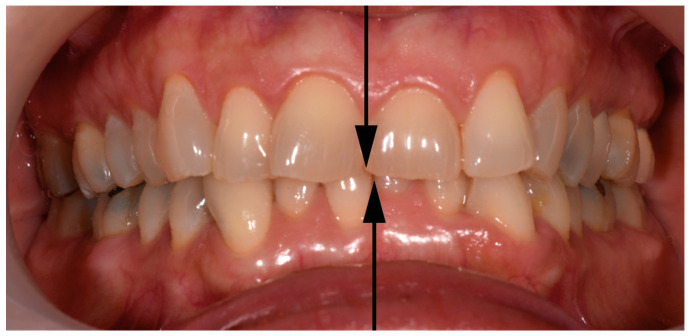
Background/Objectives: Interincisive midline deviation is frequent. Determining the cause (dental versus skeletal) is crucial for treatment planning. This study assessed the null hypothesis that neither clinical dental midline shift nor the temporomandibular disorder (TMD)-affected side correlate with maxillary/mandibular asymmetry. Methods: Thirty-eight CBCT scans were analyzed: thirty-five (92.1%) females, three (7.9%) males; mean (SD) age 34.6 (11.9) years old. Tomographic images were acquired using the i-CAT® Imaging System; mandibular/maxillary measurements were obtained with the Planmeca Romexis® software v.6.This is an ancillary study of a clinical trial (NCT02144233) that included chronic pain (TMD diagnosis; DC/TMD criteria), fully dentate, and stable normo-occlusion participants. Results: We found sixteen (42.1%) dental midline deviations to the right and thirteen (34.2%) to the left. In the study population, the right side was more developed: a hemimandible length of 119.4 (5.7) mm versus 118.6 (5.3) mm for the right and left sides, respectively (95% CI 0.21 to 1.51), p = 0.01. Conclusions: Neither the dental midline shift side nor the affected side predicted a less developed hemimandible.
DiagnosticsBiochemistry, Genetics and Molecular Biology-Clinical Biochemistry
CiteScore
4.70
自引率
8.30%
发文量
2699
审稿时长
19.64 days
期刊介绍:
Diagnostics (ISSN 2075-4418) is an international scholarly open access journal on medical diagnostics. It publishes original research articles, reviews, communications and short notes on the research and development of medical diagnostics. There is no restriction on the length of the papers. Our aim is to encourage scientists to publish their experimental and theoretical research in as much detail as possible. Full experimental and/or methodological details must be provided for research articles.





 京公网安备 11010802042870号
京ICP备2023020795号-1
京公网安备 11010802042870号
京ICP备2023020795号-1

 分享
分享
 求助内容:
求助内容: 应助结果提醒方式:
 扫码关注我们
