Vaidehi D Bhatt, Deepak C Bhatt, Kalpana D Bhatt, Supriya Dabir, Tos T J M Berendschot, Roel J Erckens, Carroll A B Webers
{"title":"“三角形”征象:一种诊断全脉络膜脱离和全脉络膜上出血的新型超声标记。","authors":"Vaidehi D Bhatt, Deepak C Bhatt, Kalpana D Bhatt, Supriya Dabir, Tos T J M Berendschot, Roel J Erckens, Carroll A B Webers","doi":"10.2147/OPTH.S495062","DOIUrl":null,"url":null,"abstract":"<p><strong>Purpose: </strong>This study aims to explore the diagnostic utility of ultrasound B-scan while introducing the \"Triangle\" sign as a novel indicator. It also validates the sign's efficacy in distinguishing between choroidal detachment (CD) and suprachoroidal hemorrhage (SCH) from retinal detachment (RD) and vitreous hemorrhage (VH).</p><p><strong>Patients and methods: </strong>Retrospective analysis of consecutive cases of total CD and SCH undergoing B-scan at a single tertiary imaging center. The study examined the presence of the \"Triangle\" sign (a hypoechoic/anechoic triangular shape of vitreous noted anterior to the optic disc) in total CD & SCH, categorized cases by subtype and etiology, and its role in differentiating from RD and VH.</p><p><strong>Results: </strong>Thirty-six eyes with a total CD and SCH were analyzed. Amongst the cases of total SCH 31 (86.1%), (58.1%) were linked to intraocular surgery, and (41.9%) were linked to post-traumatic events. The \"Triangle\" sign was consistently present in all 36 eyes, with additional findings indicating concurrent VH (52.8%) or RD (5.6%). Among the 31 eyes with total SCH, 58.3% initially had detectable choroidal membrane seen on B scan, while 41.7% did not. Despite this, the \"Triangle\" sign was consistently visible in all 36 eyes, and monitoring with B-scans revealed choroidal membrane as the hemorrhage resolved.</p><p><strong>Conclusion: </strong>The \"Triangle\" sign is a distinctive and reliable ultrasound feature for total CD and SCH diagnosis, offering clarity in challenging cases where traditional methods face limitations.</p>","PeriodicalId":93945,"journal":{"name":"Clinical ophthalmology (Auckland, N.Z.)","volume":"19 ","pages":"261-268"},"PeriodicalIF":0.0000,"publicationDate":"2025-01-22","publicationTypes":"Journal Article","fieldsOfStudy":null,"isOpenAccess":false,"openAccessPdf":"https://www.ncbi.nlm.nih.gov/pmc/articles/PMC11766375/pdf/","citationCount":"0","resultStr":"{\"title\":\"The \\\"Triangle\\\" Sign: A Novel Ultrasound Marker for Diagnosing Total Choroidal Detachment and Total Suprachoroidal Hemorrhage.\",\"authors\":\"Vaidehi D Bhatt, Deepak C Bhatt, Kalpana D Bhatt, Supriya Dabir, Tos T J M Berendschot, Roel J Erckens, Carroll A B Webers\",\"doi\":\"10.2147/OPTH.S495062\",\"DOIUrl\":null,\"url\":null,\"abstract\":\"<p><strong>Purpose: </strong>This study aims to explore the diagnostic utility of ultrasound B-scan while introducing the \\\"Triangle\\\" sign as a novel indicator. It also validates the sign's efficacy in distinguishing between choroidal detachment (CD) and suprachoroidal hemorrhage (SCH) from retinal detachment (RD) and vitreous hemorrhage (VH).</p><p><strong>Patients and methods: </strong>Retrospective analysis of consecutive cases of total CD and SCH undergoing B-scan at a single tertiary imaging center. The study examined the presence of the \\\"Triangle\\\" sign (a hypoechoic/anechoic triangular shape of vitreous noted anterior to the optic disc) in total CD & SCH, categorized cases by subtype and etiology, and its role in differentiating from RD and VH.</p><p><strong>Results: </strong>Thirty-six eyes with a total CD and SCH were analyzed. Amongst the cases of total SCH 31 (86.1%), (58.1%) were linked to intraocular surgery, and (41.9%) were linked to post-traumatic events. The \\\"Triangle\\\" sign was consistently present in all 36 eyes, with additional findings indicating concurrent VH (52.8%) or RD (5.6%). Among the 31 eyes with total SCH, 58.3% initially had detectable choroidal membrane seen on B scan, while 41.7% did not. Despite this, the \\\"Triangle\\\" sign was consistently visible in all 36 eyes, and monitoring with B-scans revealed choroidal membrane as the hemorrhage resolved.</p><p><strong>Conclusion: </strong>The \\\"Triangle\\\" sign is a distinctive and reliable ultrasound feature for total CD and SCH diagnosis, offering clarity in challenging cases where traditional methods face limitations.</p>\",\"PeriodicalId\":93945,\"journal\":{\"name\":\"Clinical ophthalmology (Auckland, N.Z.)\",\"volume\":\"19 \",\"pages\":\"261-268\"},\"PeriodicalIF\":0.0000,\"publicationDate\":\"2025-01-22\",\"publicationTypes\":\"Journal Article\",\"fieldsOfStudy\":null,\"isOpenAccess\":false,\"openAccessPdf\":\"https://www.ncbi.nlm.nih.gov/pmc/articles/PMC11766375/pdf/\",\"citationCount\":\"0\",\"resultStr\":null,\"platform\":\"Semanticscholar\",\"paperid\":null,\"PeriodicalName\":\"Clinical ophthalmology (Auckland, N.Z.)\",\"FirstCategoryId\":\"1085\",\"ListUrlMain\":\"https://doi.org/10.2147/OPTH.S495062\",\"RegionNum\":0,\"RegionCategory\":null,\"ArticlePicture\":[],\"TitleCN\":null,\"AbstractTextCN\":null,\"PMCID\":null,\"EPubDate\":\"2025/1/1 0:00:00\",\"PubModel\":\"eCollection\",\"JCR\":\"\",\"JCRName\":\"\",\"Score\":null,\"Total\":0}","platform":"Semanticscholar","paperid":null,"PeriodicalName":"Clinical ophthalmology (Auckland, N.Z.)","FirstCategoryId":"1085","ListUrlMain":"https://doi.org/10.2147/OPTH.S495062","RegionNum":0,"RegionCategory":null,"ArticlePicture":[],"TitleCN":null,"AbstractTextCN":null,"PMCID":null,"EPubDate":"2025/1/1 0:00:00","PubModel":"eCollection","JCR":"","JCRName":"","Score":null,"Total":0}
The "Triangle" Sign: A Novel Ultrasound Marker for Diagnosing Total Choroidal Detachment and Total Suprachoroidal Hemorrhage.
Purpose: This study aims to explore the diagnostic utility of ultrasound B-scan while introducing the "Triangle" sign as a novel indicator. It also validates the sign's efficacy in distinguishing between choroidal detachment (CD) and suprachoroidal hemorrhage (SCH) from retinal detachment (RD) and vitreous hemorrhage (VH).
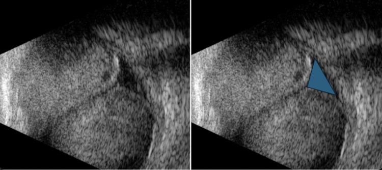
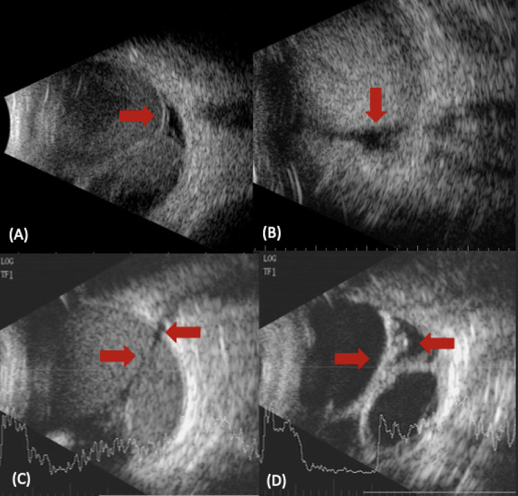
Patients and methods: Retrospective analysis of consecutive cases of total CD and SCH undergoing B-scan at a single tertiary imaging center. The study examined the presence of the "Triangle" sign (a hypoechoic/anechoic triangular shape of vitreous noted anterior to the optic disc) in total CD & SCH, categorized cases by subtype and etiology, and its role in differentiating from RD and VH.
Results: Thirty-six eyes with a total CD and SCH were analyzed. Amongst the cases of total SCH 31 (86.1%), (58.1%) were linked to intraocular surgery, and (41.9%) were linked to post-traumatic events. The "Triangle" sign was consistently present in all 36 eyes, with additional findings indicating concurrent VH (52.8%) or RD (5.6%). Among the 31 eyes with total SCH, 58.3% initially had detectable choroidal membrane seen on B scan, while 41.7% did not. Despite this, the "Triangle" sign was consistently visible in all 36 eyes, and monitoring with B-scans revealed choroidal membrane as the hemorrhage resolved.
Conclusion: The "Triangle" sign is a distinctive and reliable ultrasound feature for total CD and SCH diagnosis, offering clarity in challenging cases where traditional methods face limitations.





 京公网安备 11010802042870号
京ICP备2023020795号-1
京公网安备 11010802042870号
京ICP备2023020795号-1

 分享
分享

 求助内容:
求助内容: 应助结果提醒方式:
 扫码关注我们
