Carly Weber, Colin Wilbur, Gregg Blevins, Christian Beaulieu
{"title":"高分辨率流体抑制扩散束造影显示小儿多发性硬化症中不成比例的小穹窿和微结构改变。","authors":"Carly Weber, Colin Wilbur, Gregg Blevins, Christian Beaulieu","doi":"10.1177/20552173251315161","DOIUrl":null,"url":null,"abstract":"<p><strong>Background: </strong>Diffusion tensor imaging (DTI) in adults with multiple sclerosis (MS) has identified marked volume and diffusion abnormalities of the fornix, the main white matter (WM) output tract of the hippocampus.</p><p><strong>Objective: </strong>To determine if the fornix is affected in pediatric-onset MS (POMS) using the same DTI protocols used in adult-onset MS (AOMS), which would suggest its early involvement in the disease course.</p><p><strong>Methods: </strong>High-resolution, fluid-suppressed diffusion tractography was used to identify the fornix in 11 POMS patients (13-19 years old) and 26 controls. Fornix volume and diffusion metrics were compared between groups and with other total/regional brain volumes, and then correlated with cognitive/clinical scores.</p><p><strong>Results: </strong>POMS showed lower fornix volumes (-26%) compared to controls, which was greater than proportional losses in total and other regional brain volumes. Notably, the hippocampus volume was not lower in POMS. DTI yielded lower fractional anisotropy (-7%) and higher mean (+12%), axial (+7%), and radial (+16%) diffusivities in POMS. There were no significant correlations between fornix volume/diffusion metrics and cognitive/clinical scores.</p><p><strong>Conclusion: </strong>Diffusion tractography showed marked injury to the fornix in POMS that precedes injury to connected gray matter such as hippocampus, implicating the fornix as an early brain region affected in MS.</p>","PeriodicalId":18961,"journal":{"name":"Multiple Sclerosis Journal - Experimental, Translational and Clinical","volume":"11 1","pages":"20552173251315161"},"PeriodicalIF":2.0000,"publicationDate":"2025-01-31","publicationTypes":"Journal Article","fieldsOfStudy":null,"isOpenAccess":false,"openAccessPdf":"https://www.ncbi.nlm.nih.gov/pmc/articles/PMC11783520/pdf/","citationCount":"0","resultStr":"{\"title\":\"Disproportional smaller fornix with altered microstructure in pediatric multiple sclerosis shown by high-resolution fluid-suppressed diffusion tractography.\",\"authors\":\"Carly Weber, Colin Wilbur, Gregg Blevins, Christian Beaulieu\",\"doi\":\"10.1177/20552173251315161\",\"DOIUrl\":null,\"url\":null,\"abstract\":\"<p><strong>Background: </strong>Diffusion tensor imaging (DTI) in adults with multiple sclerosis (MS) has identified marked volume and diffusion abnormalities of the fornix, the main white matter (WM) output tract of the hippocampus.</p><p><strong>Objective: </strong>To determine if the fornix is affected in pediatric-onset MS (POMS) using the same DTI protocols used in adult-onset MS (AOMS), which would suggest its early involvement in the disease course.</p><p><strong>Methods: </strong>High-resolution, fluid-suppressed diffusion tractography was used to identify the fornix in 11 POMS patients (13-19 years old) and 26 controls. Fornix volume and diffusion metrics were compared between groups and with other total/regional brain volumes, and then correlated with cognitive/clinical scores.</p><p><strong>Results: </strong>POMS showed lower fornix volumes (-26%) compared to controls, which was greater than proportional losses in total and other regional brain volumes. Notably, the hippocampus volume was not lower in POMS. DTI yielded lower fractional anisotropy (-7%) and higher mean (+12%), axial (+7%), and radial (+16%) diffusivities in POMS. There were no significant correlations between fornix volume/diffusion metrics and cognitive/clinical scores.</p><p><strong>Conclusion: </strong>Diffusion tractography showed marked injury to the fornix in POMS that precedes injury to connected gray matter such as hippocampus, implicating the fornix as an early brain region affected in MS.</p>\",\"PeriodicalId\":18961,\"journal\":{\"name\":\"Multiple Sclerosis Journal - Experimental, Translational and Clinical\",\"volume\":\"11 1\",\"pages\":\"20552173251315161\"},\"PeriodicalIF\":2.0000,\"publicationDate\":\"2025-01-31\",\"publicationTypes\":\"Journal Article\",\"fieldsOfStudy\":null,\"isOpenAccess\":false,\"openAccessPdf\":\"https://www.ncbi.nlm.nih.gov/pmc/articles/PMC11783520/pdf/\",\"citationCount\":\"0\",\"resultStr\":null,\"platform\":\"Semanticscholar\",\"paperid\":null,\"PeriodicalName\":\"Multiple Sclerosis Journal - Experimental, Translational and Clinical\",\"FirstCategoryId\":\"1085\",\"ListUrlMain\":\"https://doi.org/10.1177/20552173251315161\",\"RegionNum\":0,\"RegionCategory\":null,\"ArticlePicture\":[],\"TitleCN\":null,\"AbstractTextCN\":null,\"PMCID\":null,\"EPubDate\":\"2025/1/1 0:00:00\",\"PubModel\":\"eCollection\",\"JCR\":\"Q2\",\"JCRName\":\"CLINICAL NEUROLOGY\",\"Score\":null,\"Total\":0}","platform":"Semanticscholar","paperid":null,"PeriodicalName":"Multiple Sclerosis Journal - Experimental, Translational and Clinical","FirstCategoryId":"1085","ListUrlMain":"https://doi.org/10.1177/20552173251315161","RegionNum":0,"RegionCategory":null,"ArticlePicture":[],"TitleCN":null,"AbstractTextCN":null,"PMCID":null,"EPubDate":"2025/1/1 0:00:00","PubModel":"eCollection","JCR":"Q2","JCRName":"CLINICAL NEUROLOGY","Score":null,"Total":0}
Disproportional smaller fornix with altered microstructure in pediatric multiple sclerosis shown by high-resolution fluid-suppressed diffusion tractography.
Background: Diffusion tensor imaging (DTI) in adults with multiple sclerosis (MS) has identified marked volume and diffusion abnormalities of the fornix, the main white matter (WM) output tract of the hippocampus.
Objective: To determine if the fornix is affected in pediatric-onset MS (POMS) using the same DTI protocols used in adult-onset MS (AOMS), which would suggest its early involvement in the disease course.
Methods: High-resolution, fluid-suppressed diffusion tractography was used to identify the fornix in 11 POMS patients (13-19 years old) and 26 controls. Fornix volume and diffusion metrics were compared between groups and with other total/regional brain volumes, and then correlated with cognitive/clinical scores.
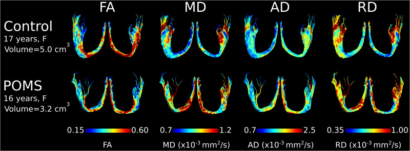
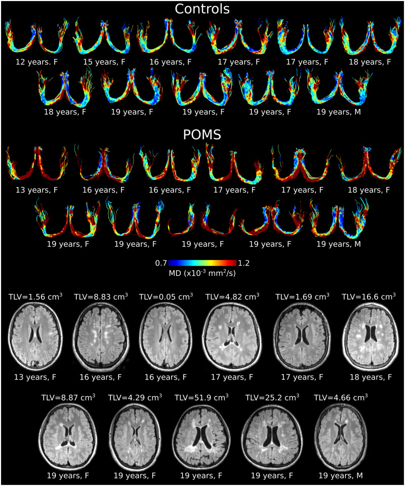
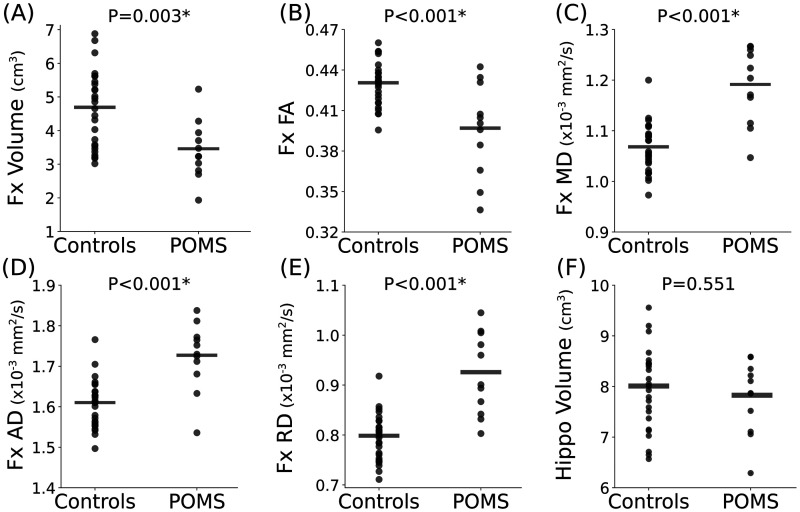
Results: POMS showed lower fornix volumes (-26%) compared to controls, which was greater than proportional losses in total and other regional brain volumes. Notably, the hippocampus volume was not lower in POMS. DTI yielded lower fractional anisotropy (-7%) and higher mean (+12%), axial (+7%), and radial (+16%) diffusivities in POMS. There were no significant correlations between fornix volume/diffusion metrics and cognitive/clinical scores.
Conclusion: Diffusion tractography showed marked injury to the fornix in POMS that precedes injury to connected gray matter such as hippocampus, implicating the fornix as an early brain region affected in MS.





 京公网安备 11010802042870号
京ICP备2023020795号-1
京公网安备 11010802042870号
京ICP备2023020795号-1

 分享
分享
 求助内容:
求助内容: 应助结果提醒方式:
 扫码关注我们
