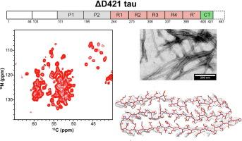
{"title":"Δ D421 截短 Tau 纤维的结构。","authors":"Nadia El Mammeri, Pu Duan, Mei Hong","doi":"10.1016/j.jmb.2025.169051","DOIUrl":null,"url":null,"abstract":"<div><div>The microtubule-associated protein tau aggregates into pathological β-sheet amyloid fibrils in Alzheimer’s disease (AD) and other neurodegenerative diseases. In these aggregates, tau is chemically modified, including abnormal hyperphosphorylation and truncation. Truncation after D421 in the C-terminal domain occurs at early stages of AD. Here we investigate the structures of ΔD421-truncated 0N4R tau fibrils assembled <em>in vitro</em> in the absence of anionic cofactors. Using solid-state NMR spectroscopy and cryoelectron microscopy, we show that ΔD421-truncated 0N4R tau forms homogeneous fibrils whose rigid core adopts a three-layered β-sheet structure that spans R2, R3 and R4 repeats. This structure is essentially identical to that of full-length tau containing phospho-mimetic mutations at the PHF1 epitope in the C-terminal domain. In comparison, a ΔD421-truncated tau that additionally contains three phospho-mimetic mutations at the AT8 epitope in the proline-rich region forms a fibril core that includes the first half of the C-terminal domain, which is excluded from all known pathological tau fibril cores. These results indicate that the posttranslational modification code of tau contains redundancy: both charge modification and truncation of the C-terminal domain promote a three-layered β-sheet structure, which resembles pathological four-repeat tau structures in several tauopathies. In comparison, reducing the positive charges at the AT8 epitope in ΔD421-truncated tau promotes a fibril core that includes an immobilized C-terminal domain. The absence of this structure in tauopathy brains implies that ΔD421 truncation does not occur in conjunction with AT8 phosphorylation in diseased brains.</div></div>","PeriodicalId":369,"journal":{"name":"Journal of Molecular Biology","volume":"437 10","pages":"Article 169051"},"PeriodicalIF":4.5000,"publicationDate":"2025-05-15","publicationTypes":"Journal Article","fieldsOfStudy":null,"isOpenAccess":false,"openAccessPdf":"","citationCount":"0","resultStr":"{\"title\":\"Structures of ΔD421 Truncated Tau Fibrils\",\"authors\":\"Nadia El Mammeri, Pu Duan, Mei Hong\",\"doi\":\"10.1016/j.jmb.2025.169051\",\"DOIUrl\":null,\"url\":null,\"abstract\":\"<div><div>The microtubule-associated protein tau aggregates into pathological β-sheet amyloid fibrils in Alzheimer’s disease (AD) and other neurodegenerative diseases. In these aggregates, tau is chemically modified, including abnormal hyperphosphorylation and truncation. Truncation after D421 in the C-terminal domain occurs at early stages of AD. Here we investigate the structures of ΔD421-truncated 0N4R tau fibrils assembled <em>in vitro</em> in the absence of anionic cofactors. Using solid-state NMR spectroscopy and cryoelectron microscopy, we show that ΔD421-truncated 0N4R tau forms homogeneous fibrils whose rigid core adopts a three-layered β-sheet structure that spans R2, R3 and R4 repeats. This structure is essentially identical to that of full-length tau containing phospho-mimetic mutations at the PHF1 epitope in the C-terminal domain. In comparison, a ΔD421-truncated tau that additionally contains three phospho-mimetic mutations at the AT8 epitope in the proline-rich region forms a fibril core that includes the first half of the C-terminal domain, which is excluded from all known pathological tau fibril cores. These results indicate that the posttranslational modification code of tau contains redundancy: both charge modification and truncation of the C-terminal domain promote a three-layered β-sheet structure, which resembles pathological four-repeat tau structures in several tauopathies. In comparison, reducing the positive charges at the AT8 epitope in ΔD421-truncated tau promotes a fibril core that includes an immobilized C-terminal domain. The absence of this structure in tauopathy brains implies that ΔD421 truncation does not occur in conjunction with AT8 phosphorylation in diseased brains.</div></div>\",\"PeriodicalId\":369,\"journal\":{\"name\":\"Journal of Molecular Biology\",\"volume\":\"437 10\",\"pages\":\"Article 169051\"},\"PeriodicalIF\":4.5000,\"publicationDate\":\"2025-05-15\",\"publicationTypes\":\"Journal Article\",\"fieldsOfStudy\":null,\"isOpenAccess\":false,\"openAccessPdf\":\"\",\"citationCount\":\"0\",\"resultStr\":null,\"platform\":\"Semanticscholar\",\"paperid\":null,\"PeriodicalName\":\"Journal of Molecular Biology\",\"FirstCategoryId\":\"99\",\"ListUrlMain\":\"https://www.sciencedirect.com/science/article/pii/S0022283625001172\",\"RegionNum\":2,\"RegionCategory\":\"生物学\",\"ArticlePicture\":[],\"TitleCN\":null,\"AbstractTextCN\":null,\"PMCID\":null,\"EPubDate\":\"2025/2/26 0:00:00\",\"PubModel\":\"Epub\",\"JCR\":\"Q1\",\"JCRName\":\"BIOCHEMISTRY & MOLECULAR BIOLOGY\",\"Score\":null,\"Total\":0}","platform":"Semanticscholar","paperid":null,"PeriodicalName":"Journal of Molecular Biology","FirstCategoryId":"99","ListUrlMain":"https://www.sciencedirect.com/science/article/pii/S0022283625001172","RegionNum":2,"RegionCategory":"生物学","ArticlePicture":[],"TitleCN":null,"AbstractTextCN":null,"PMCID":null,"EPubDate":"2025/2/26 0:00:00","PubModel":"Epub","JCR":"Q1","JCRName":"BIOCHEMISTRY & MOLECULAR BIOLOGY","Score":null,"Total":0}
The microtubule-associated protein tau aggregates into pathological β-sheet amyloid fibrils in Alzheimer’s disease (AD) and other neurodegenerative diseases. In these aggregates, tau is chemically modified, including abnormal hyperphosphorylation and truncation. Truncation after D421 in the C-terminal domain occurs at early stages of AD. Here we investigate the structures of ΔD421-truncated 0N4R tau fibrils assembled in vitro in the absence of anionic cofactors. Using solid-state NMR spectroscopy and cryoelectron microscopy, we show that ΔD421-truncated 0N4R tau forms homogeneous fibrils whose rigid core adopts a three-layered β-sheet structure that spans R2, R3 and R4 repeats. This structure is essentially identical to that of full-length tau containing phospho-mimetic mutations at the PHF1 epitope in the C-terminal domain. In comparison, a ΔD421-truncated tau that additionally contains three phospho-mimetic mutations at the AT8 epitope in the proline-rich region forms a fibril core that includes the first half of the C-terminal domain, which is excluded from all known pathological tau fibril cores. These results indicate that the posttranslational modification code of tau contains redundancy: both charge modification and truncation of the C-terminal domain promote a three-layered β-sheet structure, which resembles pathological four-repeat tau structures in several tauopathies. In comparison, reducing the positive charges at the AT8 epitope in ΔD421-truncated tau promotes a fibril core that includes an immobilized C-terminal domain. The absence of this structure in tauopathy brains implies that ΔD421 truncation does not occur in conjunction with AT8 phosphorylation in diseased brains.
期刊介绍:
Journal of Molecular Biology (JMB) provides high quality, comprehensive and broad coverage in all areas of molecular biology. The journal publishes original scientific research papers that provide mechanistic and functional insights and report a significant advance to the field. The journal encourages the submission of multidisciplinary studies that use complementary experimental and computational approaches to address challenging biological questions.
Research areas include but are not limited to: Biomolecular interactions, signaling networks, systems biology; Cell cycle, cell growth, cell differentiation; Cell death, autophagy; Cell signaling and regulation; Chemical biology; Computational biology, in combination with experimental studies; DNA replication, repair, and recombination; Development, regenerative biology, mechanistic and functional studies of stem cells; Epigenetics, chromatin structure and function; Gene expression; Membrane processes, cell surface proteins and cell-cell interactions; Methodological advances, both experimental and theoretical, including databases; Microbiology, virology, and interactions with the host or environment; Microbiota mechanistic and functional studies; Nuclear organization; Post-translational modifications, proteomics; Processing and function of biologically important macromolecules and complexes; Molecular basis of disease; RNA processing, structure and functions of non-coding RNAs, transcription; Sorting, spatiotemporal organization, trafficking; Structural biology; Synthetic biology; Translation, protein folding, chaperones, protein degradation and quality control.


 分享
分享
 求助内容:
求助内容: 应助结果提醒方式:
 扫码关注我们
