Xuejun Guo, Min Gao, Yulong Qi, Qiao Shi, Qiqiang Li, Mingmei Li, Hongbing Li
{"title":"磁共振泪囊造影对泪道及泪周病变的精确诊断。","authors":"Xuejun Guo, Min Gao, Yulong Qi, Qiao Shi, Qiqiang Li, Mingmei Li, Hongbing Li","doi":"10.1167/iovs.66.3.17","DOIUrl":null,"url":null,"abstract":"<p><strong>Purpose: </strong>The accurate determination of the etiology and location of lacrimal passage obstruction is key to treatment. However, the main assessments for lacrimal passage are invasive and have limited value. This study aimed to analyze the causes of obstruction of the lacrimal passage and investigate the diagnostic value of magnetic resonance dacryocystography (MRD).</p><p><strong>Methods: </strong>Fifty-six patients (69 eyes) with lacrimal disease underwent MRD after lacrimal irrigation. The stenotic site and degree were determined. The accuracies of the diagnoses of lesions at different sites based on MRD and routine clinical assessment were determined and compared, and a noninferiority test was used to verify the values obtained via MRD.</p><p><strong>Results: </strong>The lesion location determined through lacrimal irrigation was inconsistent with that determined through MRD. The overall accuracy of routine clinical assessment was approximately 79.7% (55 of 69) and that of MRD reached 98.6% (68 of 69). For the lesion location, the clinical misdiagnosis rates were 38.9%, 43.8%, and 63.6% for the lacrimal sac, nasolacrimal duct, and perilacrimal passage, respectively. The MRD and surgical pathology results for the lacrimal sac, nasolacrimal duct, and perilacrimal passage were consistent, except for the lacrimal canaliculus. The P value for the noninferiority of MRD to surgical pathology was 0.32.</p><p><strong>Conclusions: </strong>MRD is noninvasive and has high value for determining the cause, location, and degree of lacrimal stenosis. Its findings are nearly as accurate as those of surgical pathology. MRD should be recommended over other invasive methods for evaluating lacrimal and prelacrimal lesions.</p>","PeriodicalId":14620,"journal":{"name":"Investigative ophthalmology & visual science","volume":"66 3","pages":"17"},"PeriodicalIF":5.5000,"publicationDate":"2025-03-03","publicationTypes":"Journal Article","fieldsOfStudy":null,"isOpenAccess":false,"openAccessPdf":"https://www.ncbi.nlm.nih.gov/pmc/articles/PMC11895852/pdf/","citationCount":"0","resultStr":"{\"title\":\"Magnetic Resonance Dacryocystography for Precise Diagnosis of Lacrimal and Perilacrimal Lesions.\",\"authors\":\"Xuejun Guo, Min Gao, Yulong Qi, Qiao Shi, Qiqiang Li, Mingmei Li, Hongbing Li\",\"doi\":\"10.1167/iovs.66.3.17\",\"DOIUrl\":null,\"url\":null,\"abstract\":\"<p><strong>Purpose: </strong>The accurate determination of the etiology and location of lacrimal passage obstruction is key to treatment. However, the main assessments for lacrimal passage are invasive and have limited value. This study aimed to analyze the causes of obstruction of the lacrimal passage and investigate the diagnostic value of magnetic resonance dacryocystography (MRD).</p><p><strong>Methods: </strong>Fifty-six patients (69 eyes) with lacrimal disease underwent MRD after lacrimal irrigation. The stenotic site and degree were determined. The accuracies of the diagnoses of lesions at different sites based on MRD and routine clinical assessment were determined and compared, and a noninferiority test was used to verify the values obtained via MRD.</p><p><strong>Results: </strong>The lesion location determined through lacrimal irrigation was inconsistent with that determined through MRD. The overall accuracy of routine clinical assessment was approximately 79.7% (55 of 69) and that of MRD reached 98.6% (68 of 69). For the lesion location, the clinical misdiagnosis rates were 38.9%, 43.8%, and 63.6% for the lacrimal sac, nasolacrimal duct, and perilacrimal passage, respectively. The MRD and surgical pathology results for the lacrimal sac, nasolacrimal duct, and perilacrimal passage were consistent, except for the lacrimal canaliculus. The P value for the noninferiority of MRD to surgical pathology was 0.32.</p><p><strong>Conclusions: </strong>MRD is noninvasive and has high value for determining the cause, location, and degree of lacrimal stenosis. Its findings are nearly as accurate as those of surgical pathology. MRD should be recommended over other invasive methods for evaluating lacrimal and prelacrimal lesions.</p>\",\"PeriodicalId\":14620,\"journal\":{\"name\":\"Investigative ophthalmology & visual science\",\"volume\":\"66 3\",\"pages\":\"17\"},\"PeriodicalIF\":5.5000,\"publicationDate\":\"2025-03-03\",\"publicationTypes\":\"Journal Article\",\"fieldsOfStudy\":null,\"isOpenAccess\":false,\"openAccessPdf\":\"https://www.ncbi.nlm.nih.gov/pmc/articles/PMC11895852/pdf/\",\"citationCount\":\"0\",\"resultStr\":null,\"platform\":\"Semanticscholar\",\"paperid\":null,\"PeriodicalName\":\"Investigative ophthalmology & visual science\",\"FirstCategoryId\":\"3\",\"ListUrlMain\":\"https://doi.org/10.1167/iovs.66.3.17\",\"RegionNum\":2,\"RegionCategory\":\"医学\",\"ArticlePicture\":[],\"TitleCN\":null,\"AbstractTextCN\":null,\"PMCID\":null,\"EPubDate\":\"\",\"PubModel\":\"\",\"JCR\":\"Q1\",\"JCRName\":\"OPHTHALMOLOGY\",\"Score\":null,\"Total\":0}","platform":"Semanticscholar","paperid":null,"PeriodicalName":"Investigative ophthalmology & visual science","FirstCategoryId":"3","ListUrlMain":"https://doi.org/10.1167/iovs.66.3.17","RegionNum":2,"RegionCategory":"医学","ArticlePicture":[],"TitleCN":null,"AbstractTextCN":null,"PMCID":null,"EPubDate":"","PubModel":"","JCR":"Q1","JCRName":"OPHTHALMOLOGY","Score":null,"Total":0}
Magnetic Resonance Dacryocystography for Precise Diagnosis of Lacrimal and Perilacrimal Lesions.
Purpose: The accurate determination of the etiology and location of lacrimal passage obstruction is key to treatment. However, the main assessments for lacrimal passage are invasive and have limited value. This study aimed to analyze the causes of obstruction of the lacrimal passage and investigate the diagnostic value of magnetic resonance dacryocystography (MRD).
Methods: Fifty-six patients (69 eyes) with lacrimal disease underwent MRD after lacrimal irrigation. The stenotic site and degree were determined. The accuracies of the diagnoses of lesions at different sites based on MRD and routine clinical assessment were determined and compared, and a noninferiority test was used to verify the values obtained via MRD.
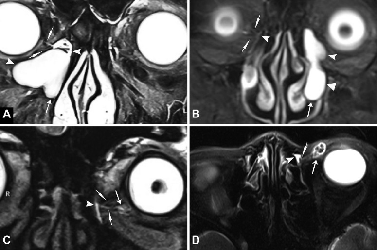
Results: The lesion location determined through lacrimal irrigation was inconsistent with that determined through MRD. The overall accuracy of routine clinical assessment was approximately 79.7% (55 of 69) and that of MRD reached 98.6% (68 of 69). For the lesion location, the clinical misdiagnosis rates were 38.9%, 43.8%, and 63.6% for the lacrimal sac, nasolacrimal duct, and perilacrimal passage, respectively. The MRD and surgical pathology results for the lacrimal sac, nasolacrimal duct, and perilacrimal passage were consistent, except for the lacrimal canaliculus. The P value for the noninferiority of MRD to surgical pathology was 0.32.
Conclusions: MRD is noninvasive and has high value for determining the cause, location, and degree of lacrimal stenosis. Its findings are nearly as accurate as those of surgical pathology. MRD should be recommended over other invasive methods for evaluating lacrimal and prelacrimal lesions.
期刊介绍:
Investigative Ophthalmology & Visual Science (IOVS), published as ready online, is a peer-reviewed academic journal of the Association for Research in Vision and Ophthalmology (ARVO). IOVS features original research, mostly pertaining to clinical and laboratory ophthalmology and vision research in general.






 京公网安备 11010802042870号
京ICP备2023020795号-1
京公网安备 11010802042870号
京ICP备2023020795号-1

 分享
分享
 求助内容:
求助内容: 应助结果提醒方式:
 扫码关注我们
