Samantha Lee, Robert Chen, Sumeet Kumar, Weiling Lee, Huihua Li, Louis C S Tan, Eng King Tan, Nicole C H Keong, Ling Ling Chan
{"title":"脾角轴位T1和FLAIR MRI图像与特发性正常压力脑积水。","authors":"Samantha Lee, Robert Chen, Sumeet Kumar, Weiling Lee, Huihua Li, Louis C S Tan, Eng King Tan, Nicole C H Keong, Ling Ling Chan","doi":"10.1093/bjr/tqaf058","DOIUrl":null,"url":null,"abstract":"<p><strong>Objectives: </strong>The splenial angle (SA), measured on axial DTI colour fractional anisotropy MRI, outperformed the callosal angle (CA) in predicting idiopathic normal pressure hydrocephalus (NPH) patients from those with Alzheimer's dementia, Parkinson's disease (PD), and healthy controls (HC). We investigated its reliability and classification performance on more commonly acquired T1 magnetization-prepared rapid gradient-echo (MPRAGE) and fluid-attenuated inversion-recovery (FLAIR) images.</p><p><strong>Methods: </strong>Splenial angle was measured on axial MPRAGE and FLAIR images in 57 subjects (19 NPH, PD and HC each) by 2 raters and compared across groups. Receiver operating characteristics (ROC) analysis was used to assess its classification performance, differentiating NPH from non-NPH groups, in comparison to the CA.</p><p><strong>Results: </strong>Inter-rater reliability for SA was excellent (intraclass correlation coefficients ≥ 0.91). Splenial angle was effective in differentiating NPH from non-NPH patients on MPRAGE and FLAIR images (P < .001). Its ROC curves showed excellent performance classifying NPH from HC (Area under the curve [AUC] 1) and PD (AUC > 0.93) groups and were highly comparable to those for CA (1; 0.947). Angles wider than 60° and narrower than 45° robustly (100%) excluded and predicted NPH from HC, respectively. The narrower 45° cutoff yielded better sensitivity (84.2%-89.5%) in differentiating NPH from PD patients.</p><p><strong>Conclusions: </strong>The SA on MPRAGE/FLAIR images showed excellent inter-rater reliability and classification performance predicting NPH from non-NPH groups, rivalling those of the CA.</p><p><strong>Advances in knowledge: </strong>The SA on MPRAGE and FLAIR images is reproducible and shows excellent diagnostic performance, differentiating NPH from non-NPH groups, with potential to replace the CA in NPH screening given its accessibility on routine axial neuroimaging.</p>","PeriodicalId":9306,"journal":{"name":"British Journal of Radiology","volume":" ","pages":"938-946"},"PeriodicalIF":3.4000,"publicationDate":"2025-06-01","publicationTypes":"Journal Article","fieldsOfStudy":null,"isOpenAccess":false,"openAccessPdf":"https://www.ncbi.nlm.nih.gov/pmc/articles/PMC12089770/pdf/","citationCount":"0","resultStr":"{\"title\":\"Splenial angle on axial T1 and fluid-attenuated inversion-recovery MRI images and idiopathic normal pressure hydrocephalus.\",\"authors\":\"Samantha Lee, Robert Chen, Sumeet Kumar, Weiling Lee, Huihua Li, Louis C S Tan, Eng King Tan, Nicole C H Keong, Ling Ling Chan\",\"doi\":\"10.1093/bjr/tqaf058\",\"DOIUrl\":null,\"url\":null,\"abstract\":\"<p><strong>Objectives: </strong>The splenial angle (SA), measured on axial DTI colour fractional anisotropy MRI, outperformed the callosal angle (CA) in predicting idiopathic normal pressure hydrocephalus (NPH) patients from those with Alzheimer's dementia, Parkinson's disease (PD), and healthy controls (HC). We investigated its reliability and classification performance on more commonly acquired T1 magnetization-prepared rapid gradient-echo (MPRAGE) and fluid-attenuated inversion-recovery (FLAIR) images.</p><p><strong>Methods: </strong>Splenial angle was measured on axial MPRAGE and FLAIR images in 57 subjects (19 NPH, PD and HC each) by 2 raters and compared across groups. Receiver operating characteristics (ROC) analysis was used to assess its classification performance, differentiating NPH from non-NPH groups, in comparison to the CA.</p><p><strong>Results: </strong>Inter-rater reliability for SA was excellent (intraclass correlation coefficients ≥ 0.91). Splenial angle was effective in differentiating NPH from non-NPH patients on MPRAGE and FLAIR images (P < .001). Its ROC curves showed excellent performance classifying NPH from HC (Area under the curve [AUC] 1) and PD (AUC > 0.93) groups and were highly comparable to those for CA (1; 0.947). Angles wider than 60° and narrower than 45° robustly (100%) excluded and predicted NPH from HC, respectively. The narrower 45° cutoff yielded better sensitivity (84.2%-89.5%) in differentiating NPH from PD patients.</p><p><strong>Conclusions: </strong>The SA on MPRAGE/FLAIR images showed excellent inter-rater reliability and classification performance predicting NPH from non-NPH groups, rivalling those of the CA.</p><p><strong>Advances in knowledge: </strong>The SA on MPRAGE and FLAIR images is reproducible and shows excellent diagnostic performance, differentiating NPH from non-NPH groups, with potential to replace the CA in NPH screening given its accessibility on routine axial neuroimaging.</p>\",\"PeriodicalId\":9306,\"journal\":{\"name\":\"British Journal of Radiology\",\"volume\":\" \",\"pages\":\"938-946\"},\"PeriodicalIF\":3.4000,\"publicationDate\":\"2025-06-01\",\"publicationTypes\":\"Journal Article\",\"fieldsOfStudy\":null,\"isOpenAccess\":false,\"openAccessPdf\":\"https://www.ncbi.nlm.nih.gov/pmc/articles/PMC12089770/pdf/\",\"citationCount\":\"0\",\"resultStr\":null,\"platform\":\"Semanticscholar\",\"paperid\":null,\"PeriodicalName\":\"British Journal of Radiology\",\"FirstCategoryId\":\"3\",\"ListUrlMain\":\"https://doi.org/10.1093/bjr/tqaf058\",\"RegionNum\":4,\"RegionCategory\":\"医学\",\"ArticlePicture\":[],\"TitleCN\":null,\"AbstractTextCN\":null,\"PMCID\":null,\"EPubDate\":\"\",\"PubModel\":\"\",\"JCR\":\"Q3\",\"JCRName\":\"RADIOLOGY, NUCLEAR MEDICINE & MEDICAL IMAGING\",\"Score\":null,\"Total\":0}","platform":"Semanticscholar","paperid":null,"PeriodicalName":"British Journal of Radiology","FirstCategoryId":"3","ListUrlMain":"https://doi.org/10.1093/bjr/tqaf058","RegionNum":4,"RegionCategory":"医学","ArticlePicture":[],"TitleCN":null,"AbstractTextCN":null,"PMCID":null,"EPubDate":"","PubModel":"","JCR":"Q3","JCRName":"RADIOLOGY, NUCLEAR MEDICINE & MEDICAL IMAGING","Score":null,"Total":0}
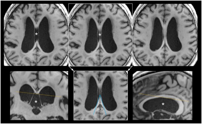
Splenial angle on axial T1 and fluid-attenuated inversion-recovery MRI images and idiopathic normal pressure hydrocephalus.
Objectives: The splenial angle (SA), measured on axial DTI colour fractional anisotropy MRI, outperformed the callosal angle (CA) in predicting idiopathic normal pressure hydrocephalus (NPH) patients from those with Alzheimer's dementia, Parkinson's disease (PD), and healthy controls (HC). We investigated its reliability and classification performance on more commonly acquired T1 magnetization-prepared rapid gradient-echo (MPRAGE) and fluid-attenuated inversion-recovery (FLAIR) images.
Methods: Splenial angle was measured on axial MPRAGE and FLAIR images in 57 subjects (19 NPH, PD and HC each) by 2 raters and compared across groups. Receiver operating characteristics (ROC) analysis was used to assess its classification performance, differentiating NPH from non-NPH groups, in comparison to the CA.
Results: Inter-rater reliability for SA was excellent (intraclass correlation coefficients ≥ 0.91). Splenial angle was effective in differentiating NPH from non-NPH patients on MPRAGE and FLAIR images (P < .001). Its ROC curves showed excellent performance classifying NPH from HC (Area under the curve [AUC] 1) and PD (AUC > 0.93) groups and were highly comparable to those for CA (1; 0.947). Angles wider than 60° and narrower than 45° robustly (100%) excluded and predicted NPH from HC, respectively. The narrower 45° cutoff yielded better sensitivity (84.2%-89.5%) in differentiating NPH from PD patients.
Conclusions: The SA on MPRAGE/FLAIR images showed excellent inter-rater reliability and classification performance predicting NPH from non-NPH groups, rivalling those of the CA.
Advances in knowledge: The SA on MPRAGE and FLAIR images is reproducible and shows excellent diagnostic performance, differentiating NPH from non-NPH groups, with potential to replace the CA in NPH screening given its accessibility on routine axial neuroimaging.
期刊介绍:
BJR is the international research journal of the British Institute of Radiology and is the oldest scientific journal in the field of radiology and related sciences.
Dating back to 1896, BJR’s history is radiology’s history, and the journal has featured some landmark papers such as the first description of Computed Tomography "Computerized transverse axial tomography" by Godfrey Hounsfield in 1973. A valuable historical resource, the complete BJR archive has been digitized from 1896.
Quick Facts:
- 2015 Impact Factor – 1.840
- Receipt to first decision – average of 6 weeks
- Acceptance to online publication – average of 3 weeks
- ISSN: 0007-1285
- eISSN: 1748-880X
Open Access option






 京公网安备 11010802042870号
京ICP备2023020795号-1
京公网安备 11010802042870号
京ICP备2023020795号-1

 分享
分享
 求助内容:
求助内容: 应助结果提醒方式:
 扫码关注我们
