Maryam Alkadhimi, Anuradha Helen Manne, Yanyan Jiang, Marcus Green, Anderson Joseph Ryan
{"title":"用于研究肺部药物辐射反应的器官型模型。","authors":"Maryam Alkadhimi, Anuradha Helen Manne, Yanyan Jiang, Marcus Green, Anderson Joseph Ryan","doi":"10.14440/jbm.2025.0080","DOIUrl":null,"url":null,"abstract":"<p><strong>Background: </strong>Established <i>in vivo</i> radiobiological models are commonly used to assess anti-tumor effects and normal tissue toxicity. However, these models have notable limitations, and additional models are necessary to gain a deeper insights into drug-radiation interactions.</p><p><strong>Objective: </strong>This study aimed to develop an organotypic <i>ex vivo</i> model by using precision-cut lung slices (PCLSs) to evaluate radiation-induced residual deoxyribonucleic acid (DNA) damage, both alone and in combination with a pharmacological inhibitor of DNA double-strand break (DSB) repair.</p><p><strong>Methods: </strong>Left lungs from female C57BL/6 mice were dissected, perfused with 4% low-gelling-temperature agarose, and sliced into 250 μm sections. Lung slices were then incubated <i>ex vivo</i> for up to 7 days. The slices were irradiated using <sup>137</sup>Cs, either with or without a DNA-dependent protein kinase (DNA-PK) inhibitor (NU7441). Tissue sections were subsequently fixed and stained for γH2AX and 53BP1, which serve as histological markers of DNA DSBs.</p><p><strong>Results: </strong>The established conditions preserved tissue viability for up to 7 days and maintained structural integrity for 2 days. DNA damage, detected through γH2AX and 53BP1 staining, was consistent between lungs irradiated <i>ex vivo</i> and their counterparts irradiated <i>in vivo</i>. In the organotypic model, radiation alone in DNA-PK-deficient SCID mice and radiation combined with DNA-PK inhibition in C57BL/6 mice led to increased residual γH2AX and 53BP1 staining.</p><p><strong>Conclusion: </strong>This study demonstrates that residual DNA damage levels following ionizing radiation in lung tissue are comparable between <i>in vivo</i> and <i>ex vivo</i> tissue slices, suggesting that PCLSs serve as a valuable organotypic model for investigating the effects of drug-radiation combinations.</p>","PeriodicalId":73618,"journal":{"name":"Journal of biological methods","volume":"12 1","pages":"e99010041"},"PeriodicalIF":0.0000,"publicationDate":"2024-11-28","publicationTypes":"Journal Article","fieldsOfStudy":null,"isOpenAccess":false,"openAccessPdf":"https://www.ncbi.nlm.nih.gov/pmc/articles/PMC11973049/pdf/","citationCount":"0","resultStr":"{\"title\":\"An organotypic model for investigating drug-radiation responses in the lung.\",\"authors\":\"Maryam Alkadhimi, Anuradha Helen Manne, Yanyan Jiang, Marcus Green, Anderson Joseph Ryan\",\"doi\":\"10.14440/jbm.2025.0080\",\"DOIUrl\":null,\"url\":null,\"abstract\":\"<p><strong>Background: </strong>Established <i>in vivo</i> radiobiological models are commonly used to assess anti-tumor effects and normal tissue toxicity. However, these models have notable limitations, and additional models are necessary to gain a deeper insights into drug-radiation interactions.</p><p><strong>Objective: </strong>This study aimed to develop an organotypic <i>ex vivo</i> model by using precision-cut lung slices (PCLSs) to evaluate radiation-induced residual deoxyribonucleic acid (DNA) damage, both alone and in combination with a pharmacological inhibitor of DNA double-strand break (DSB) repair.</p><p><strong>Methods: </strong>Left lungs from female C57BL/6 mice were dissected, perfused with 4% low-gelling-temperature agarose, and sliced into 250 μm sections. Lung slices were then incubated <i>ex vivo</i> for up to 7 days. The slices were irradiated using <sup>137</sup>Cs, either with or without a DNA-dependent protein kinase (DNA-PK) inhibitor (NU7441). Tissue sections were subsequently fixed and stained for γH2AX and 53BP1, which serve as histological markers of DNA DSBs.</p><p><strong>Results: </strong>The established conditions preserved tissue viability for up to 7 days and maintained structural integrity for 2 days. DNA damage, detected through γH2AX and 53BP1 staining, was consistent between lungs irradiated <i>ex vivo</i> and their counterparts irradiated <i>in vivo</i>. In the organotypic model, radiation alone in DNA-PK-deficient SCID mice and radiation combined with DNA-PK inhibition in C57BL/6 mice led to increased residual γH2AX and 53BP1 staining.</p><p><strong>Conclusion: </strong>This study demonstrates that residual DNA damage levels following ionizing radiation in lung tissue are comparable between <i>in vivo</i> and <i>ex vivo</i> tissue slices, suggesting that PCLSs serve as a valuable organotypic model for investigating the effects of drug-radiation combinations.</p>\",\"PeriodicalId\":73618,\"journal\":{\"name\":\"Journal of biological methods\",\"volume\":\"12 1\",\"pages\":\"e99010041\"},\"PeriodicalIF\":0.0000,\"publicationDate\":\"2024-11-28\",\"publicationTypes\":\"Journal Article\",\"fieldsOfStudy\":null,\"isOpenAccess\":false,\"openAccessPdf\":\"https://www.ncbi.nlm.nih.gov/pmc/articles/PMC11973049/pdf/\",\"citationCount\":\"0\",\"resultStr\":null,\"platform\":\"Semanticscholar\",\"paperid\":null,\"PeriodicalName\":\"Journal of biological methods\",\"FirstCategoryId\":\"1085\",\"ListUrlMain\":\"https://doi.org/10.14440/jbm.2025.0080\",\"RegionNum\":0,\"RegionCategory\":null,\"ArticlePicture\":[],\"TitleCN\":null,\"AbstractTextCN\":null,\"PMCID\":null,\"EPubDate\":\"2025/1/1 0:00:00\",\"PubModel\":\"eCollection\",\"JCR\":\"\",\"JCRName\":\"\",\"Score\":null,\"Total\":0}","platform":"Semanticscholar","paperid":null,"PeriodicalName":"Journal of biological methods","FirstCategoryId":"1085","ListUrlMain":"https://doi.org/10.14440/jbm.2025.0080","RegionNum":0,"RegionCategory":null,"ArticlePicture":[],"TitleCN":null,"AbstractTextCN":null,"PMCID":null,"EPubDate":"2025/1/1 0:00:00","PubModel":"eCollection","JCR":"","JCRName":"","Score":null,"Total":0}
An organotypic model for investigating drug-radiation responses in the lung.
Background: Established in vivo radiobiological models are commonly used to assess anti-tumor effects and normal tissue toxicity. However, these models have notable limitations, and additional models are necessary to gain a deeper insights into drug-radiation interactions.
Objective: This study aimed to develop an organotypic ex vivo model by using precision-cut lung slices (PCLSs) to evaluate radiation-induced residual deoxyribonucleic acid (DNA) damage, both alone and in combination with a pharmacological inhibitor of DNA double-strand break (DSB) repair.
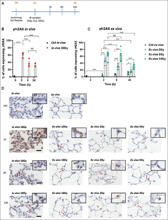
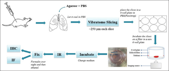
Methods: Left lungs from female C57BL/6 mice were dissected, perfused with 4% low-gelling-temperature agarose, and sliced into 250 μm sections. Lung slices were then incubated ex vivo for up to 7 days. The slices were irradiated using 137Cs, either with or without a DNA-dependent protein kinase (DNA-PK) inhibitor (NU7441). Tissue sections were subsequently fixed and stained for γH2AX and 53BP1, which serve as histological markers of DNA DSBs.
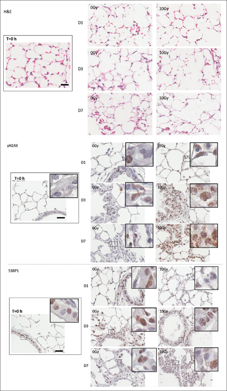
Results: The established conditions preserved tissue viability for up to 7 days and maintained structural integrity for 2 days. DNA damage, detected through γH2AX and 53BP1 staining, was consistent between lungs irradiated ex vivo and their counterparts irradiated in vivo. In the organotypic model, radiation alone in DNA-PK-deficient SCID mice and radiation combined with DNA-PK inhibition in C57BL/6 mice led to increased residual γH2AX and 53BP1 staining.
Conclusion: This study demonstrates that residual DNA damage levels following ionizing radiation in lung tissue are comparable between in vivo and ex vivo tissue slices, suggesting that PCLSs serve as a valuable organotypic model for investigating the effects of drug-radiation combinations.


 分享
分享

 求助内容:
求助内容: 应助结果提醒方式:
 扫码关注我们
