J Jacquemier, JP Molès, F Penault-Llorca, J Adélaide, M Torrente, P Viens, D Birnbaum, C Theillet
{"title":"用四种单克隆抗体对乳腺癌中的 p53 进行免疫组化分析:染色结果与 PCR-SSCP 结果的比较","authors":"J Jacquemier, JP Molès, F Penault-Llorca, J Adélaide, M Torrente, P Viens, D Birnbaum, C Theillet","doi":"10.1038/bjc.1994.164","DOIUrl":null,"url":null,"abstract":"The expression of p53 protein was examined in a series of 136 primary breast carcinomas, 106 of which were analysed with a panel of four monoclonal antibodies (MAbs 1801, 240, DO7 and DO1). p53 expression was detected with at least one antibody in 40 tumours (38%), whereas only 15 tumours (14%) were positive with all four antibodies. Some variability in the immunostaining could be observed depending on the antibody used. This was noticeable both for the number of positive cells within a section and for the intensity of staining. We therefore selected a panel of 17 tumour sections (nine were highly positive, three with medium to low staining and five with low to negative staining), which we analysed by polymerase chain reaction-single-strand conformation polymorphism analysis (PCR-SSCP) for the presence of a p53 mutation at the molecular level. Mutations were identified in 15 cases. Therefore the proportion of p53-stained cells does not seem to be an exact representation of the number of cancer cells bearing a mutation within a tumour. A statistically significant correlation was observed between p53 expression, regardless of the number of positive antibodies, and grade III disease (P < 0.0001), oestrogen (P < 0.0001) or progesterone receptor negativity (P = 0.0061), increased Ki 67 index (P = 0.0018), epidermal growth factor receptor (EGFR) positivity (P = 0.0076) and aneuploidy (P = 0.037). No correlation was observed with tumour size or lymph node involvement. In univariate analysis p53 expression was not correlated with disease-free survival, in contrast to the classical prognostic parameters, which were statistically correlated. In this series p53 expression was not a marker of early recurrence.","PeriodicalId":9243,"journal":{"name":"British Journal of Cancer","volume":"69 5","pages":"846-852"},"PeriodicalIF":6.8000,"publicationDate":"1994-05-01","publicationTypes":"Journal Article","fieldsOfStudy":null,"isOpenAccess":false,"openAccessPdf":"https://sci-hub-pdf.com/10.1038/bjc.1994.164","citationCount":"111","resultStr":"{\"title\":\"p53 immunohistochemical analysis in breast cancer with four monoclonal antibodies: comparison of staining and PCR-SSCP results\",\"authors\":\"J Jacquemier, JP Molès, F Penault-Llorca, J Adélaide, M Torrente, P Viens, D Birnbaum, C Theillet\",\"doi\":\"10.1038/bjc.1994.164\",\"DOIUrl\":null,\"url\":null,\"abstract\":\"The expression of p53 protein was examined in a series of 136 primary breast carcinomas, 106 of which were analysed with a panel of four monoclonal antibodies (MAbs 1801, 240, DO7 and DO1). p53 expression was detected with at least one antibody in 40 tumours (38%), whereas only 15 tumours (14%) were positive with all four antibodies. Some variability in the immunostaining could be observed depending on the antibody used. This was noticeable both for the number of positive cells within a section and for the intensity of staining. We therefore selected a panel of 17 tumour sections (nine were highly positive, three with medium to low staining and five with low to negative staining), which we analysed by polymerase chain reaction-single-strand conformation polymorphism analysis (PCR-SSCP) for the presence of a p53 mutation at the molecular level. Mutations were identified in 15 cases. Therefore the proportion of p53-stained cells does not seem to be an exact representation of the number of cancer cells bearing a mutation within a tumour. A statistically significant correlation was observed between p53 expression, regardless of the number of positive antibodies, and grade III disease (P < 0.0001), oestrogen (P < 0.0001) or progesterone receptor negativity (P = 0.0061), increased Ki 67 index (P = 0.0018), epidermal growth factor receptor (EGFR) positivity (P = 0.0076) and aneuploidy (P = 0.037). No correlation was observed with tumour size or lymph node involvement. In univariate analysis p53 expression was not correlated with disease-free survival, in contrast to the classical prognostic parameters, which were statistically correlated. In this series p53 expression was not a marker of early recurrence.\",\"PeriodicalId\":9243,\"journal\":{\"name\":\"British Journal of Cancer\",\"volume\":\"69 5\",\"pages\":\"846-852\"},\"PeriodicalIF\":6.8000,\"publicationDate\":\"1994-05-01\",\"publicationTypes\":\"Journal Article\",\"fieldsOfStudy\":null,\"isOpenAccess\":false,\"openAccessPdf\":\"https://sci-hub-pdf.com/10.1038/bjc.1994.164\",\"citationCount\":\"111\",\"resultStr\":null,\"platform\":\"Semanticscholar\",\"paperid\":null,\"PeriodicalName\":\"British Journal of Cancer\",\"FirstCategoryId\":\"3\",\"ListUrlMain\":\"https://www.nature.com/articles/bjc1994164\",\"RegionNum\":1,\"RegionCategory\":\"医学\",\"ArticlePicture\":[],\"TitleCN\":null,\"AbstractTextCN\":null,\"PMCID\":null,\"EPubDate\":\"\",\"PubModel\":\"\",\"JCR\":\"Q1\",\"JCRName\":\"ONCOLOGY\",\"Score\":null,\"Total\":0}","platform":"Semanticscholar","paperid":null,"PeriodicalName":"British Journal of Cancer","FirstCategoryId":"3","ListUrlMain":"https://www.nature.com/articles/bjc1994164","RegionNum":1,"RegionCategory":"医学","ArticlePicture":[],"TitleCN":null,"AbstractTextCN":null,"PMCID":null,"EPubDate":"","PubModel":"","JCR":"Q1","JCRName":"ONCOLOGY","Score":null,"Total":0}
p53 immunohistochemical analysis in breast cancer with four monoclonal antibodies: comparison of staining and PCR-SSCP results
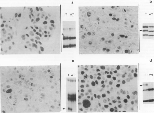
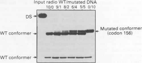
The expression of p53 protein was examined in a series of 136 primary breast carcinomas, 106 of which were analysed with a panel of four monoclonal antibodies (MAbs 1801, 240, DO7 and DO1). p53 expression was detected with at least one antibody in 40 tumours (38%), whereas only 15 tumours (14%) were positive with all four antibodies. Some variability in the immunostaining could be observed depending on the antibody used. This was noticeable both for the number of positive cells within a section and for the intensity of staining. We therefore selected a panel of 17 tumour sections (nine were highly positive, three with medium to low staining and five with low to negative staining), which we analysed by polymerase chain reaction-single-strand conformation polymorphism analysis (PCR-SSCP) for the presence of a p53 mutation at the molecular level. Mutations were identified in 15 cases. Therefore the proportion of p53-stained cells does not seem to be an exact representation of the number of cancer cells bearing a mutation within a tumour. A statistically significant correlation was observed between p53 expression, regardless of the number of positive antibodies, and grade III disease (P < 0.0001), oestrogen (P < 0.0001) or progesterone receptor negativity (P = 0.0061), increased Ki 67 index (P = 0.0018), epidermal growth factor receptor (EGFR) positivity (P = 0.0076) and aneuploidy (P = 0.037). No correlation was observed with tumour size or lymph node involvement. In univariate analysis p53 expression was not correlated with disease-free survival, in contrast to the classical prognostic parameters, which were statistically correlated. In this series p53 expression was not a marker of early recurrence.
期刊介绍:
The British Journal of Cancer is one of the most-cited general cancer journals, publishing significant advances in translational and clinical cancer research.It also publishes high-quality reviews and thought-provoking comment on all aspects of cancer prevention,diagnosis and treatment.


 分享
分享
 求助内容:
求助内容: 应助结果提醒方式:
 扫码关注我们
