Sugandha Goel, Purna Nangia, A Joash Rijey, Kumar Saurabh, Rupak Roy
{"title":"不寻常的黄斑毛细血管扩张2型伴大量色素沉积。","authors":"Sugandha Goel, Purna Nangia, A Joash Rijey, Kumar Saurabh, Rupak Roy","doi":"10.3205/oc000176","DOIUrl":null,"url":null,"abstract":"Macular telangiectasia type 2 (MacTel) is a bilateral retinal disease that seems to be limited to the juxtafoveal region of the macula. We herein report an unusual presentation of MacTel with a large pigment deposit at the macula. Fundus of the right eye showed a large pigment deposit at the macula and right-angled venule. The left eye fundus showed a grayish discoloration due to subretinal fibrosis, dark pigment clumps and right-angled venule in the macula. Lesions were highlighted on multicolor imaging and blue reflectance imaging. Spectral domain optical coherence tomography (SD-OCT) of both eyes showed hyperreflectivity on the inner aspect of the retina corresponding to the area of pigment clumping.","PeriodicalId":73178,"journal":{"name":"GMS ophthalmology cases","volume":"11 ","pages":"Doc03"},"PeriodicalIF":0.0000,"publicationDate":"2021-01-28","publicationTypes":"Journal Article","fieldsOfStudy":null,"isOpenAccess":false,"openAccessPdf":"https://www.ncbi.nlm.nih.gov/pmc/articles/PMC7894179/pdf/","citationCount":"0","resultStr":"{\"title\":\"An unusual presentation of macular telangiectasia type 2 with a large pigment deposit.\",\"authors\":\"Sugandha Goel, Purna Nangia, A Joash Rijey, Kumar Saurabh, Rupak Roy\",\"doi\":\"10.3205/oc000176\",\"DOIUrl\":null,\"url\":null,\"abstract\":\"Macular telangiectasia type 2 (MacTel) is a bilateral retinal disease that seems to be limited to the juxtafoveal region of the macula. We herein report an unusual presentation of MacTel with a large pigment deposit at the macula. Fundus of the right eye showed a large pigment deposit at the macula and right-angled venule. The left eye fundus showed a grayish discoloration due to subretinal fibrosis, dark pigment clumps and right-angled venule in the macula. Lesions were highlighted on multicolor imaging and blue reflectance imaging. Spectral domain optical coherence tomography (SD-OCT) of both eyes showed hyperreflectivity on the inner aspect of the retina corresponding to the area of pigment clumping.\",\"PeriodicalId\":73178,\"journal\":{\"name\":\"GMS ophthalmology cases\",\"volume\":\"11 \",\"pages\":\"Doc03\"},\"PeriodicalIF\":0.0000,\"publicationDate\":\"2021-01-28\",\"publicationTypes\":\"Journal Article\",\"fieldsOfStudy\":null,\"isOpenAccess\":false,\"openAccessPdf\":\"https://www.ncbi.nlm.nih.gov/pmc/articles/PMC7894179/pdf/\",\"citationCount\":\"0\",\"resultStr\":null,\"platform\":\"Semanticscholar\",\"paperid\":null,\"PeriodicalName\":\"GMS ophthalmology cases\",\"FirstCategoryId\":\"1085\",\"ListUrlMain\":\"https://doi.org/10.3205/oc000176\",\"RegionNum\":0,\"RegionCategory\":null,\"ArticlePicture\":[],\"TitleCN\":null,\"AbstractTextCN\":null,\"PMCID\":null,\"EPubDate\":\"2021/1/1 0:00:00\",\"PubModel\":\"eCollection\",\"JCR\":\"\",\"JCRName\":\"\",\"Score\":null,\"Total\":0}","platform":"Semanticscholar","paperid":null,"PeriodicalName":"GMS ophthalmology cases","FirstCategoryId":"1085","ListUrlMain":"https://doi.org/10.3205/oc000176","RegionNum":0,"RegionCategory":null,"ArticlePicture":[],"TitleCN":null,"AbstractTextCN":null,"PMCID":null,"EPubDate":"2021/1/1 0:00:00","PubModel":"eCollection","JCR":"","JCRName":"","Score":null,"Total":0}
An unusual presentation of macular telangiectasia type 2 with a large pigment deposit.
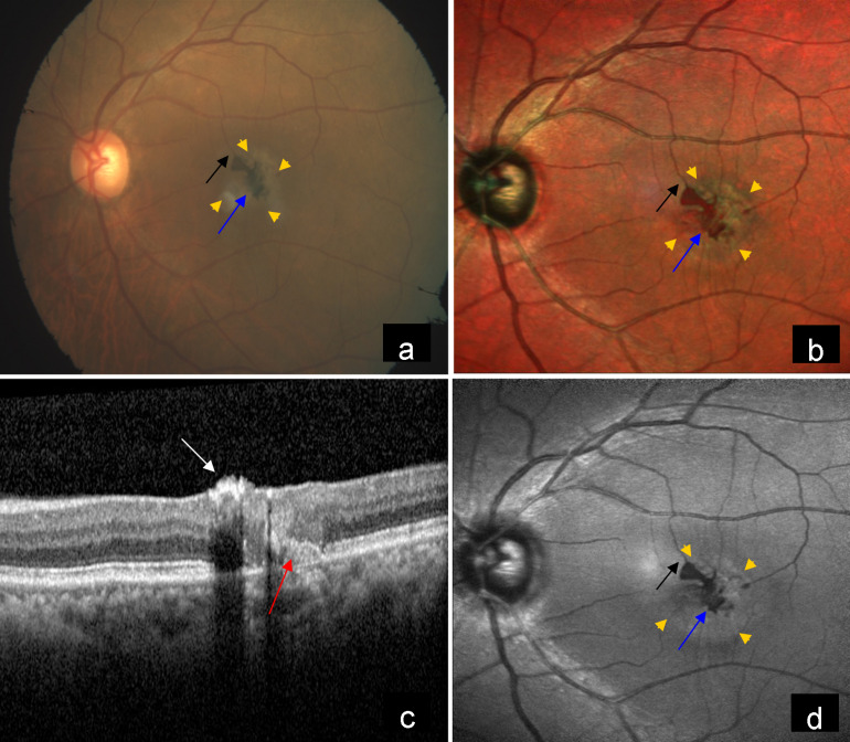
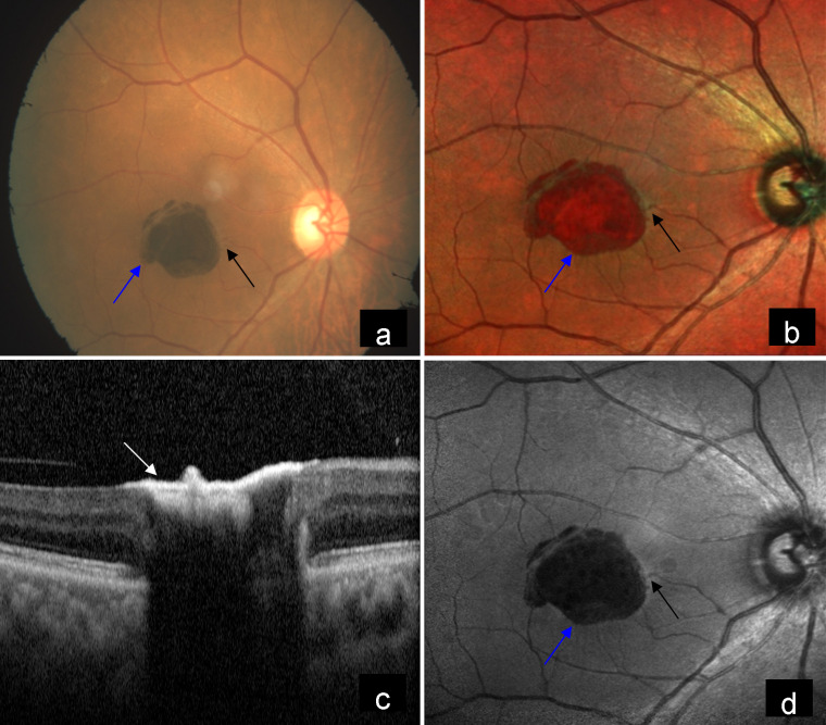
Macular telangiectasia type 2 (MacTel) is a bilateral retinal disease that seems to be limited to the juxtafoveal region of the macula. We herein report an unusual presentation of MacTel with a large pigment deposit at the macula. Fundus of the right eye showed a large pigment deposit at the macula and right-angled venule. The left eye fundus showed a grayish discoloration due to subretinal fibrosis, dark pigment clumps and right-angled venule in the macula. Lesions were highlighted on multicolor imaging and blue reflectance imaging. Spectral domain optical coherence tomography (SD-OCT) of both eyes showed hyperreflectivity on the inner aspect of the retina corresponding to the area of pigment clumping.


 分享
分享

 求助内容:
求助内容: 应助结果提醒方式:
 扫码关注我们
