Enrique Arciniegas, Luz Marina Carrillo, Héctor Rojas, Jacinto Pineda, Richard Ramírez, Oscar Reyes, Marina Chopite, Albani Rocheta
{"title":"丰满的内皮细胞与先前存在的静脉整合,促进了脓性肉芽肿中 \"母 \"血管和 \"子 \"血管的形成:galectin-1、-3 和 -8 的可能作用。","authors":"Enrique Arciniegas, Luz Marina Carrillo, Héctor Rojas, Jacinto Pineda, Richard Ramírez, Oscar Reyes, Marina Chopite, Albani Rocheta","doi":"10.1177/2059513120986687","DOIUrl":null,"url":null,"abstract":"<p><strong>Introduction: </strong>Pyogenic granuloma (PG) is a reactive inflammatory vascular lesion of the skin and mucous membranes, characterised by the presence of enlarged venules and seamed and seamless capillaries with plump endothelial cells (EC), and numerous macrophages. EC activation upregulates the synthesis of galectins and induces their translocation to the EC surface promoting angiogenesis and lymphangiogenesis, particularly galectin-1 (Gal-1), Gal-3 and Gal-8. However, the presence and distribution of Gal-1, -3 and -8, as well as their implications in the pathogenesis of PG, has not been considered.</p><p><strong>Materials and methods: </strong>Eight biopsies from patients diagnosed with PG were selected. The presence of PECAM-1/CD31, IL-1β, VEGF-C, VEGFR-2, VEGFR-3, integrin β1, CD44, fibronectin and Gal-1, -3 and -8 was assessed by immunofluorescence staining using confocal laser scanning microscopy.</p><p><strong>Results and discussion: </strong>Immunostaining revealed that these molecules were present in the enlarged venules with plump ECs, in some macrophages and other immune cells. We propose that macrophages release VEGF-A and VEGF-C inducing VEGFR-2/VEGFR-3 expression and activation, leading macrophages to transdifferentiate into plump ECs that might integrate into pre-existing venules, contributing to the formation of enlarged venules with transluminal bridges and capillaries. EC activation, induced by certain cytokines, has been shown to stimulate galectin expression and changes in the cellular localisation through association and activation of specific EC surface glycoproteins. Therefore, it is plausible that Gal-1, -3 and -8, acting in a concerted manner, could be mediating the transdifferentiation of macrophages into plump ECs and facilitating their migration and incorporation into the new vessels.</p><p><strong>Lay summary: </strong>In this study, immunostaining of pyogenic granuloma (PG) tissue sections showed immunoreactivity for PECAM-1/CD31, IL-1β, VEGF-C, VEGFR-2 and VEGFR-3, and galectin-1, -3 and -8 in enlarged venules with plump endothelial cells (EC), as well as in some macrophages and other immune cells. Interestingly, enlarged and thin-walled transient vessels lined by PECAM-1/CD31 and VEGFR-2 immunopositive ECs that form from pre-existing normal venules in response to VEGF-A (called 'mother' vessels [MV]) and that undergo intraluminal bridging evolving into various types of capillaries (called 'daughter' vessels [DV]) have been observed in benign and malignant tumours, in physiological and pathological angiogenesis as well as in vascular malformations, suggesting an important role for VEGF-A and VEGFR-2 in such a process. However, it is not only the mechanisms by which the MVs evolve in different types of DVs that remains to be elucidated, but also whether the cells that form intraluminal bridges proceed from locally activated ECs or whether they are derived from bone marrow precursors or from resident macrophages.Given that the formation of homodimers by Gal-1 and Gal-8 and pentamers by Gal-3 to generate gal-glycan lattices at the cell surface and in the extracellular space has been shown, it is possible that in PG tissue Gal-1, -3 and -8, through their binding partners, form a supramolecular structure at the surface of ECs and plump ECs, macrophages and in the extracellular space that might be mediating the transdifferentiation of macrophages into plump ECs and facilitating the migration and incorporation of these cells into the pre-existing venules, thus contributing to the formation of MVs and DVs.</p>","PeriodicalId":21495,"journal":{"name":"Scars, burns & healing","volume":"7 ","pages":"2059513120986687"},"PeriodicalIF":0.0000,"publicationDate":"2021-01-22","publicationTypes":"Journal Article","fieldsOfStudy":null,"isOpenAccess":false,"openAccessPdf":"https://ftp.ncbi.nlm.nih.gov/pub/pmc/oa_pdf/48/cf/10.1177_2059513120986687.PMC7841855.pdf","citationCount":"0","resultStr":"{\"title\":\"Plump endothelial cells integrated into pre-existing venules contribute to the formation of 'mother' and 'daughter' vessels in pyogenic granuloma: possible role of galectin-1, -3 and -8.\",\"authors\":\"Enrique Arciniegas, Luz Marina Carrillo, Héctor Rojas, Jacinto Pineda, Richard Ramírez, Oscar Reyes, Marina Chopite, Albani Rocheta\",\"doi\":\"10.1177/2059513120986687\",\"DOIUrl\":null,\"url\":null,\"abstract\":\"<p><strong>Introduction: </strong>Pyogenic granuloma (PG) is a reactive inflammatory vascular lesion of the skin and mucous membranes, characterised by the presence of enlarged venules and seamed and seamless capillaries with plump endothelial cells (EC), and numerous macrophages. EC activation upregulates the synthesis of galectins and induces their translocation to the EC surface promoting angiogenesis and lymphangiogenesis, particularly galectin-1 (Gal-1), Gal-3 and Gal-8. However, the presence and distribution of Gal-1, -3 and -8, as well as their implications in the pathogenesis of PG, has not been considered.</p><p><strong>Materials and methods: </strong>Eight biopsies from patients diagnosed with PG were selected. The presence of PECAM-1/CD31, IL-1β, VEGF-C, VEGFR-2, VEGFR-3, integrin β1, CD44, fibronectin and Gal-1, -3 and -8 was assessed by immunofluorescence staining using confocal laser scanning microscopy.</p><p><strong>Results and discussion: </strong>Immunostaining revealed that these molecules were present in the enlarged venules with plump ECs, in some macrophages and other immune cells. We propose that macrophages release VEGF-A and VEGF-C inducing VEGFR-2/VEGFR-3 expression and activation, leading macrophages to transdifferentiate into plump ECs that might integrate into pre-existing venules, contributing to the formation of enlarged venules with transluminal bridges and capillaries. EC activation, induced by certain cytokines, has been shown to stimulate galectin expression and changes in the cellular localisation through association and activation of specific EC surface glycoproteins. Therefore, it is plausible that Gal-1, -3 and -8, acting in a concerted manner, could be mediating the transdifferentiation of macrophages into plump ECs and facilitating their migration and incorporation into the new vessels.</p><p><strong>Lay summary: </strong>In this study, immunostaining of pyogenic granuloma (PG) tissue sections showed immunoreactivity for PECAM-1/CD31, IL-1β, VEGF-C, VEGFR-2 and VEGFR-3, and galectin-1, -3 and -8 in enlarged venules with plump endothelial cells (EC), as well as in some macrophages and other immune cells. Interestingly, enlarged and thin-walled transient vessels lined by PECAM-1/CD31 and VEGFR-2 immunopositive ECs that form from pre-existing normal venules in response to VEGF-A (called 'mother' vessels [MV]) and that undergo intraluminal bridging evolving into various types of capillaries (called 'daughter' vessels [DV]) have been observed in benign and malignant tumours, in physiological and pathological angiogenesis as well as in vascular malformations, suggesting an important role for VEGF-A and VEGFR-2 in such a process. However, it is not only the mechanisms by which the MVs evolve in different types of DVs that remains to be elucidated, but also whether the cells that form intraluminal bridges proceed from locally activated ECs or whether they are derived from bone marrow precursors or from resident macrophages.Given that the formation of homodimers by Gal-1 and Gal-8 and pentamers by Gal-3 to generate gal-glycan lattices at the cell surface and in the extracellular space has been shown, it is possible that in PG tissue Gal-1, -3 and -8, through their binding partners, form a supramolecular structure at the surface of ECs and plump ECs, macrophages and in the extracellular space that might be mediating the transdifferentiation of macrophages into plump ECs and facilitating the migration and incorporation of these cells into the pre-existing venules, thus contributing to the formation of MVs and DVs.</p>\",\"PeriodicalId\":21495,\"journal\":{\"name\":\"Scars, burns & healing\",\"volume\":\"7 \",\"pages\":\"2059513120986687\"},\"PeriodicalIF\":0.0000,\"publicationDate\":\"2021-01-22\",\"publicationTypes\":\"Journal Article\",\"fieldsOfStudy\":null,\"isOpenAccess\":false,\"openAccessPdf\":\"https://ftp.ncbi.nlm.nih.gov/pub/pmc/oa_pdf/48/cf/10.1177_2059513120986687.PMC7841855.pdf\",\"citationCount\":\"0\",\"resultStr\":null,\"platform\":\"Semanticscholar\",\"paperid\":null,\"PeriodicalName\":\"Scars, burns & healing\",\"FirstCategoryId\":\"1085\",\"ListUrlMain\":\"https://doi.org/10.1177/2059513120986687\",\"RegionNum\":0,\"RegionCategory\":null,\"ArticlePicture\":[],\"TitleCN\":null,\"AbstractTextCN\":null,\"PMCID\":null,\"EPubDate\":\"2021/1/1 0:00:00\",\"PubModel\":\"eCollection\",\"JCR\":\"\",\"JCRName\":\"\",\"Score\":null,\"Total\":0}","platform":"Semanticscholar","paperid":null,"PeriodicalName":"Scars, burns & healing","FirstCategoryId":"1085","ListUrlMain":"https://doi.org/10.1177/2059513120986687","RegionNum":0,"RegionCategory":null,"ArticlePicture":[],"TitleCN":null,"AbstractTextCN":null,"PMCID":null,"EPubDate":"2021/1/1 0:00:00","PubModel":"eCollection","JCR":"","JCRName":"","Score":null,"Total":0}
Plump endothelial cells integrated into pre-existing venules contribute to the formation of 'mother' and 'daughter' vessels in pyogenic granuloma: possible role of galectin-1, -3 and -8.
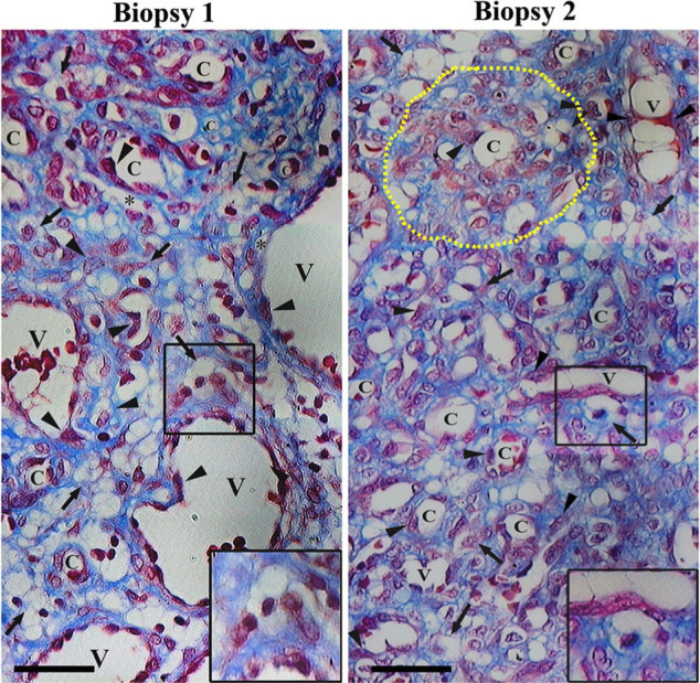
Introduction: Pyogenic granuloma (PG) is a reactive inflammatory vascular lesion of the skin and mucous membranes, characterised by the presence of enlarged venules and seamed and seamless capillaries with plump endothelial cells (EC), and numerous macrophages. EC activation upregulates the synthesis of galectins and induces their translocation to the EC surface promoting angiogenesis and lymphangiogenesis, particularly galectin-1 (Gal-1), Gal-3 and Gal-8. However, the presence and distribution of Gal-1, -3 and -8, as well as their implications in the pathogenesis of PG, has not been considered.
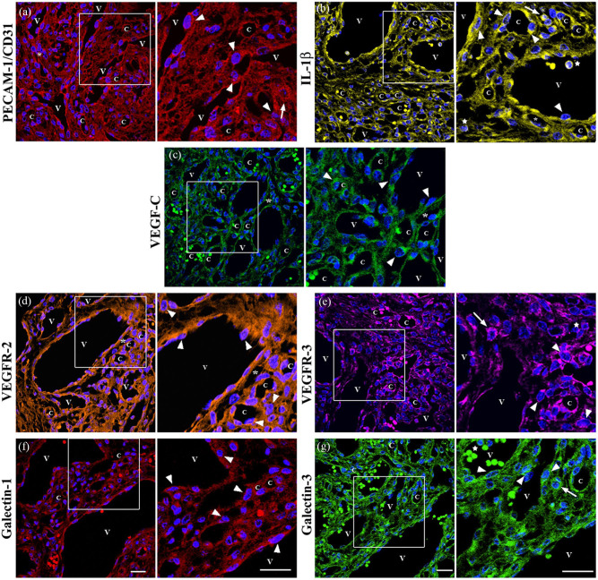
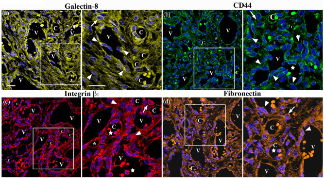
Materials and methods: Eight biopsies from patients diagnosed with PG were selected. The presence of PECAM-1/CD31, IL-1β, VEGF-C, VEGFR-2, VEGFR-3, integrin β1, CD44, fibronectin and Gal-1, -3 and -8 was assessed by immunofluorescence staining using confocal laser scanning microscopy.
Results and discussion: Immunostaining revealed that these molecules were present in the enlarged venules with plump ECs, in some macrophages and other immune cells. We propose that macrophages release VEGF-A and VEGF-C inducing VEGFR-2/VEGFR-3 expression and activation, leading macrophages to transdifferentiate into plump ECs that might integrate into pre-existing venules, contributing to the formation of enlarged venules with transluminal bridges and capillaries. EC activation, induced by certain cytokines, has been shown to stimulate galectin expression and changes in the cellular localisation through association and activation of specific EC surface glycoproteins. Therefore, it is plausible that Gal-1, -3 and -8, acting in a concerted manner, could be mediating the transdifferentiation of macrophages into plump ECs and facilitating their migration and incorporation into the new vessels.
Lay summary: In this study, immunostaining of pyogenic granuloma (PG) tissue sections showed immunoreactivity for PECAM-1/CD31, IL-1β, VEGF-C, VEGFR-2 and VEGFR-3, and galectin-1, -3 and -8 in enlarged venules with plump endothelial cells (EC), as well as in some macrophages and other immune cells. Interestingly, enlarged and thin-walled transient vessels lined by PECAM-1/CD31 and VEGFR-2 immunopositive ECs that form from pre-existing normal venules in response to VEGF-A (called 'mother' vessels [MV]) and that undergo intraluminal bridging evolving into various types of capillaries (called 'daughter' vessels [DV]) have been observed in benign and malignant tumours, in physiological and pathological angiogenesis as well as in vascular malformations, suggesting an important role for VEGF-A and VEGFR-2 in such a process. However, it is not only the mechanisms by which the MVs evolve in different types of DVs that remains to be elucidated, but also whether the cells that form intraluminal bridges proceed from locally activated ECs or whether they are derived from bone marrow precursors or from resident macrophages.Given that the formation of homodimers by Gal-1 and Gal-8 and pentamers by Gal-3 to generate gal-glycan lattices at the cell surface and in the extracellular space has been shown, it is possible that in PG tissue Gal-1, -3 and -8, through their binding partners, form a supramolecular structure at the surface of ECs and plump ECs, macrophages and in the extracellular space that might be mediating the transdifferentiation of macrophages into plump ECs and facilitating the migration and incorporation of these cells into the pre-existing venules, thus contributing to the formation of MVs and DVs.


 分享
分享

 求助内容:
求助内容: 应助结果提醒方式:
 扫码关注我们
