{"title":"胶原酶在体内促进细胞对损伤和伤口愈合的反应。","authors":"Kathleen N Riley, Ira M Herman","doi":"","DOIUrl":null,"url":null,"abstract":"<p><strong>Objective: </strong>This study focuses on the growth-promoting and migration-enhancing role that Clostridial collagenase plays in vitro and in vivo.</p><p><strong>Methods: </strong>For in vitro studies, biosynthesized extracellular matrices were treated with purified Clostridial collagenase, nonspecific proteases, or buffer controls. Keratinocytes were subsequently plated upon these matrices in the presence or absence of Clostridial collagenase and/or heparin-binding epidermal-like growth factor, and cell proliferation and migration were quantified. To examine the effects of Clostridial collagenase in vivo, we performed a double-blind study of full-thickness wounds on the backs of Yucatan Micropigs, testing the effects of purified Clostridial collagenase, Regranex (PDGF-BB), and Solosite (carboxymethyl cellulose) on wound healing.</p><p><strong>Results: </strong></p><p><strong>In vitro studies: </strong>Matrix pretreatment with Clostridial collagenase stimulates a 2-fold increase in proliferation and postinjury migration; when Clostridial collagenase and/or heparin-binding epidermal-like growth factor are added to the growth media, there is an additional doubling of growth and migration, yielding approximately 5-fold enhancement of keratinocyte proliferation and migration. Papain-urea treatment under similar conditions results in a 50% decrease in cell number over a 1-week time course. In vivo studies: By all parameters measured, including granulation tissue formation, inflammation, re-epithelization, and time to wound closure, purified Clostridial collagenase was superior (analysis of variance, P > .05) to other treatments tested.</p><p><strong>Conclusion: </strong>On the basis of these findings, we concluded that Clostridial collagenase stimulates keratinocyte cellular responses to injury in vitro and may represent a novel therapeutic approach for promotion of wound healing in vivo.</p>","PeriodicalId":87438,"journal":{"name":"Journal of burns and wounds","volume":"4 ","pages":"e8"},"PeriodicalIF":0.0000,"publicationDate":"2005-05-17","publicationTypes":"Journal Article","fieldsOfStudy":null,"isOpenAccess":false,"openAccessPdf":"https://www.ncbi.nlm.nih.gov/pmc/articles/PMC1501117/pdf/","citationCount":"0","resultStr":"{\"title\":\"Collagenase promotes the cellular responses to injury and wound healing in vivo.\",\"authors\":\"Kathleen N Riley, Ira M Herman\",\"doi\":\"\",\"DOIUrl\":null,\"url\":null,\"abstract\":\"<p><strong>Objective: </strong>This study focuses on the growth-promoting and migration-enhancing role that Clostridial collagenase plays in vitro and in vivo.</p><p><strong>Methods: </strong>For in vitro studies, biosynthesized extracellular matrices were treated with purified Clostridial collagenase, nonspecific proteases, or buffer controls. Keratinocytes were subsequently plated upon these matrices in the presence or absence of Clostridial collagenase and/or heparin-binding epidermal-like growth factor, and cell proliferation and migration were quantified. To examine the effects of Clostridial collagenase in vivo, we performed a double-blind study of full-thickness wounds on the backs of Yucatan Micropigs, testing the effects of purified Clostridial collagenase, Regranex (PDGF-BB), and Solosite (carboxymethyl cellulose) on wound healing.</p><p><strong>Results: </strong></p><p><strong>In vitro studies: </strong>Matrix pretreatment with Clostridial collagenase stimulates a 2-fold increase in proliferation and postinjury migration; when Clostridial collagenase and/or heparin-binding epidermal-like growth factor are added to the growth media, there is an additional doubling of growth and migration, yielding approximately 5-fold enhancement of keratinocyte proliferation and migration. Papain-urea treatment under similar conditions results in a 50% decrease in cell number over a 1-week time course. In vivo studies: By all parameters measured, including granulation tissue formation, inflammation, re-epithelization, and time to wound closure, purified Clostridial collagenase was superior (analysis of variance, P > .05) to other treatments tested.</p><p><strong>Conclusion: </strong>On the basis of these findings, we concluded that Clostridial collagenase stimulates keratinocyte cellular responses to injury in vitro and may represent a novel therapeutic approach for promotion of wound healing in vivo.</p>\",\"PeriodicalId\":87438,\"journal\":{\"name\":\"Journal of burns and wounds\",\"volume\":\"4 \",\"pages\":\"e8\"},\"PeriodicalIF\":0.0000,\"publicationDate\":\"2005-05-17\",\"publicationTypes\":\"Journal Article\",\"fieldsOfStudy\":null,\"isOpenAccess\":false,\"openAccessPdf\":\"https://www.ncbi.nlm.nih.gov/pmc/articles/PMC1501117/pdf/\",\"citationCount\":\"0\",\"resultStr\":null,\"platform\":\"Semanticscholar\",\"paperid\":null,\"PeriodicalName\":\"Journal of burns and wounds\",\"FirstCategoryId\":\"1085\",\"ListUrlMain\":\"\",\"RegionNum\":0,\"RegionCategory\":null,\"ArticlePicture\":[],\"TitleCN\":null,\"AbstractTextCN\":null,\"PMCID\":null,\"EPubDate\":\"\",\"PubModel\":\"\",\"JCR\":\"\",\"JCRName\":\"\",\"Score\":null,\"Total\":0}","platform":"Semanticscholar","paperid":null,"PeriodicalName":"Journal of burns and wounds","FirstCategoryId":"1085","ListUrlMain":"","RegionNum":0,"RegionCategory":null,"ArticlePicture":[],"TitleCN":null,"AbstractTextCN":null,"PMCID":null,"EPubDate":"","PubModel":"","JCR":"","JCRName":"","Score":null,"Total":0}
Collagenase promotes the cellular responses to injury and wound healing in vivo.
Objective: This study focuses on the growth-promoting and migration-enhancing role that Clostridial collagenase plays in vitro and in vivo.
Methods: For in vitro studies, biosynthesized extracellular matrices were treated with purified Clostridial collagenase, nonspecific proteases, or buffer controls. Keratinocytes were subsequently plated upon these matrices in the presence or absence of Clostridial collagenase and/or heparin-binding epidermal-like growth factor, and cell proliferation and migration were quantified. To examine the effects of Clostridial collagenase in vivo, we performed a double-blind study of full-thickness wounds on the backs of Yucatan Micropigs, testing the effects of purified Clostridial collagenase, Regranex (PDGF-BB), and Solosite (carboxymethyl cellulose) on wound healing.
Results:
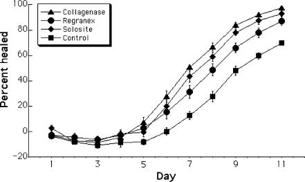
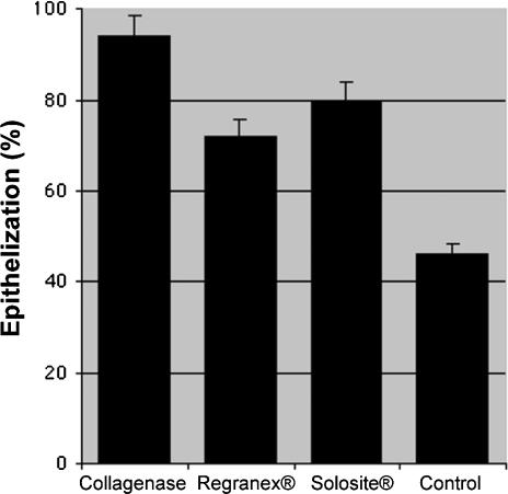
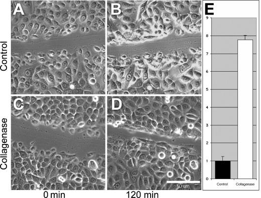
In vitro studies: Matrix pretreatment with Clostridial collagenase stimulates a 2-fold increase in proliferation and postinjury migration; when Clostridial collagenase and/or heparin-binding epidermal-like growth factor are added to the growth media, there is an additional doubling of growth and migration, yielding approximately 5-fold enhancement of keratinocyte proliferation and migration. Papain-urea treatment under similar conditions results in a 50% decrease in cell number over a 1-week time course. In vivo studies: By all parameters measured, including granulation tissue formation, inflammation, re-epithelization, and time to wound closure, purified Clostridial collagenase was superior (analysis of variance, P > .05) to other treatments tested.
Conclusion: On the basis of these findings, we concluded that Clostridial collagenase stimulates keratinocyte cellular responses to injury in vitro and may represent a novel therapeutic approach for promotion of wound healing in vivo.
 分享
分享


 求助内容:
求助内容: 应助结果提醒方式:
 扫码关注我们
