Pavel Komínek, Ivo Stárek, Marie Geierová, Petr Matoušek, Karol Zeleník
{"title":"鼻窦区磷化间充质瘤:1例报告及文献复习。","authors":"Pavel Komínek, Ivo Stárek, Marie Geierová, Petr Matoušek, Karol Zeleník","doi":"10.1186/1758-3284-3-16","DOIUrl":null,"url":null,"abstract":"<p><strong>Background: </strong>Oncogenous osteomalacia (OOM), which is also known as tumour-induced osteomalacia, is a rare condition associated with a neoplasm and a related systemic bone demineralization caused by renal phosphate wasting. OOM usually occurs in association with a variety of different mesenchymal tumours, and they were categorized into four distinct morphological patterns which they termed \"phosphaturic mesenchymal tumour\". Of its 4 histopathological subtypes, the mixed connective tissue variant is most commonly observed. Only 10% of cases appear in the head and neck regions and moreover, only 5 previously published tumors were localized in the sinonasal area. The authors describe a case of a man with a PMT originating from the frontoethmoidal region.</p><p><strong>Case presentation: </strong>A 53-year-old man was referred to our ORL clinic due to a presence of a mass at the nasal root having been growing asymptomatically for 1 year. CT scans demonstrated a large (25 × 20 × 35 mm) bilateral frontoethmoidal mass with destruction of nasal bones. The tumor did not appear to invade to the anterior skull base. A selective angiography revealed a moderate hypervascularization of the tumour during early and late arterial phases. The tumour was removed from the external approach and the definitive histopathological diagnosis was a phospaturic mesenchymal tumor. Dual energy X-ray absorptiometry revealed a slight osteopenia of the first and second lumbar vertebrae and neck of the thigh bone. The serum and urinary levels of both calcium and anorganic phosphate were within normal limits. The patient is doing well three years after the operation, and the serum and urine levels of calcium and phosphate remain well within normal limits.</p><p><strong>Conclusion: </strong>PMT is rare in the sinonasal region, it can be rarely observed without the signs of osteomalacia.</p>","PeriodicalId":49195,"journal":{"name":"Head and Neck Optical Diagnostics Society","volume":"3 ","pages":"16"},"PeriodicalIF":0.0000,"publicationDate":"2011-03-16","publicationTypes":"Journal Article","fieldsOfStudy":null,"isOpenAccess":false,"openAccessPdf":"https://sci-hub-pdf.com/10.1186/1758-3284-3-16","citationCount":"21","resultStr":"{\"title\":\"Phosphaturic mesenchymal tumour of the sinonasal area: case report and review of the literature.\",\"authors\":\"Pavel Komínek, Ivo Stárek, Marie Geierová, Petr Matoušek, Karol Zeleník\",\"doi\":\"10.1186/1758-3284-3-16\",\"DOIUrl\":null,\"url\":null,\"abstract\":\"<p><strong>Background: </strong>Oncogenous osteomalacia (OOM), which is also known as tumour-induced osteomalacia, is a rare condition associated with a neoplasm and a related systemic bone demineralization caused by renal phosphate wasting. OOM usually occurs in association with a variety of different mesenchymal tumours, and they were categorized into four distinct morphological patterns which they termed \\\"phosphaturic mesenchymal tumour\\\". Of its 4 histopathological subtypes, the mixed connective tissue variant is most commonly observed. Only 10% of cases appear in the head and neck regions and moreover, only 5 previously published tumors were localized in the sinonasal area. The authors describe a case of a man with a PMT originating from the frontoethmoidal region.</p><p><strong>Case presentation: </strong>A 53-year-old man was referred to our ORL clinic due to a presence of a mass at the nasal root having been growing asymptomatically for 1 year. CT scans demonstrated a large (25 × 20 × 35 mm) bilateral frontoethmoidal mass with destruction of nasal bones. The tumor did not appear to invade to the anterior skull base. A selective angiography revealed a moderate hypervascularization of the tumour during early and late arterial phases. The tumour was removed from the external approach and the definitive histopathological diagnosis was a phospaturic mesenchymal tumor. Dual energy X-ray absorptiometry revealed a slight osteopenia of the first and second lumbar vertebrae and neck of the thigh bone. The serum and urinary levels of both calcium and anorganic phosphate were within normal limits. The patient is doing well three years after the operation, and the serum and urine levels of calcium and phosphate remain well within normal limits.</p><p><strong>Conclusion: </strong>PMT is rare in the sinonasal region, it can be rarely observed without the signs of osteomalacia.</p>\",\"PeriodicalId\":49195,\"journal\":{\"name\":\"Head and Neck Optical Diagnostics Society\",\"volume\":\"3 \",\"pages\":\"16\"},\"PeriodicalIF\":0.0000,\"publicationDate\":\"2011-03-16\",\"publicationTypes\":\"Journal Article\",\"fieldsOfStudy\":null,\"isOpenAccess\":false,\"openAccessPdf\":\"https://sci-hub-pdf.com/10.1186/1758-3284-3-16\",\"citationCount\":\"21\",\"resultStr\":null,\"platform\":\"Semanticscholar\",\"paperid\":null,\"PeriodicalName\":\"Head and Neck Optical Diagnostics Society\",\"FirstCategoryId\":\"1085\",\"ListUrlMain\":\"https://doi.org/10.1186/1758-3284-3-16\",\"RegionNum\":0,\"RegionCategory\":null,\"ArticlePicture\":[],\"TitleCN\":null,\"AbstractTextCN\":null,\"PMCID\":null,\"EPubDate\":\"\",\"PubModel\":\"\",\"JCR\":\"\",\"JCRName\":\"\",\"Score\":null,\"Total\":0}","platform":"Semanticscholar","paperid":null,"PeriodicalName":"Head and Neck Optical Diagnostics Society","FirstCategoryId":"1085","ListUrlMain":"https://doi.org/10.1186/1758-3284-3-16","RegionNum":0,"RegionCategory":null,"ArticlePicture":[],"TitleCN":null,"AbstractTextCN":null,"PMCID":null,"EPubDate":"","PubModel":"","JCR":"","JCRName":"","Score":null,"Total":0}
Phosphaturic mesenchymal tumour of the sinonasal area: case report and review of the literature.
Background: Oncogenous osteomalacia (OOM), which is also known as tumour-induced osteomalacia, is a rare condition associated with a neoplasm and a related systemic bone demineralization caused by renal phosphate wasting. OOM usually occurs in association with a variety of different mesenchymal tumours, and they were categorized into four distinct morphological patterns which they termed "phosphaturic mesenchymal tumour". Of its 4 histopathological subtypes, the mixed connective tissue variant is most commonly observed. Only 10% of cases appear in the head and neck regions and moreover, only 5 previously published tumors were localized in the sinonasal area. The authors describe a case of a man with a PMT originating from the frontoethmoidal region.
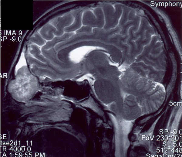
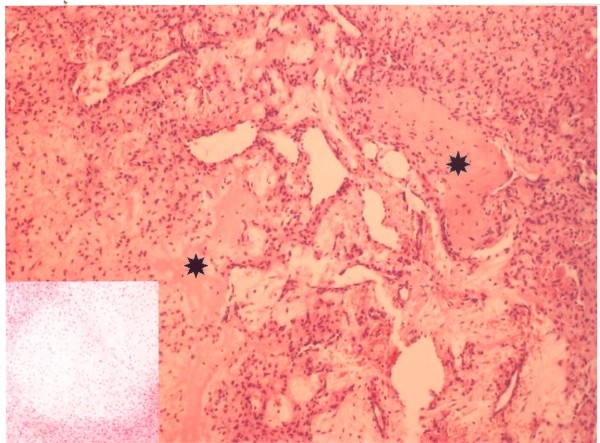
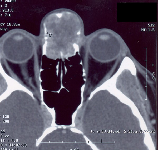
Case presentation: A 53-year-old man was referred to our ORL clinic due to a presence of a mass at the nasal root having been growing asymptomatically for 1 year. CT scans demonstrated a large (25 × 20 × 35 mm) bilateral frontoethmoidal mass with destruction of nasal bones. The tumor did not appear to invade to the anterior skull base. A selective angiography revealed a moderate hypervascularization of the tumour during early and late arterial phases. The tumour was removed from the external approach and the definitive histopathological diagnosis was a phospaturic mesenchymal tumor. Dual energy X-ray absorptiometry revealed a slight osteopenia of the first and second lumbar vertebrae and neck of the thigh bone. The serum and urinary levels of both calcium and anorganic phosphate were within normal limits. The patient is doing well three years after the operation, and the serum and urine levels of calcium and phosphate remain well within normal limits.
Conclusion: PMT is rare in the sinonasal region, it can be rarely observed without the signs of osteomalacia.


 分享
分享

 求助内容:
求助内容: 应助结果提醒方式:
 扫码关注我们
