Valentina Rivelli, Heinz T Luebbers, Franz E Weber, Claudia Cordella, Klaus W Grätz, Astrid L Kruse
{"title":"筛查头颈癌复发及淋巴结转移:计算机断层扫描在随访中的作用。","authors":"Valentina Rivelli, Heinz T Luebbers, Franz E Weber, Claudia Cordella, Klaus W Grätz, Astrid L Kruse","doi":"10.1186/1758-3284-3-18","DOIUrl":null,"url":null,"abstract":"<p><strong>Introduction: </strong>Follow-up of patients with oral cancer is being questioned with regard to financial costs and effectiveness. Therefore, the aim of the present study was to evaluate whether local recurrence and cervical lymph node metastases were first discovered clinically or by routine computer tomography.</p><p><strong>Materials and methods: </strong>The records of all 317 patients that were treated for an oral cancer between 1998 and 2008 were systematically reviewed. Criteria for inclusion were tumor histology with a squamous cell carcinoma of the head and neck, and regular follow-up examinations with a minimum follow-up time of 12 months, including clinical and radiological (CT) controls. All patients had the first CT after 6 months, followed by yearly CT controls.</p><p><strong>Results: </strong>Out of 315 patients with an oral squamous cell carcinoma, 294 were evaluated. Those experiencing neither recurrence of the tumor nor lymph node metastases constituted 62%. Local recurrence was seen in 36 (12%), lymph node metastases in 32 (11%), and both in 16 (6%). Of the 32 patients with lymph node metastases, 25 were recognized first clinically, and 7 were detected by routine CT scans; concerning local recurrence, 32 appeared clinically, and 4 were detected by routine CT scans.</p><p><strong>Conclusion: </strong>Routine CT for follow-up is still indicated for detecting lymph node metastases as well as local recurrence.</p>","PeriodicalId":49195,"journal":{"name":"Head and Neck Optical Diagnostics Society","volume":"3 ","pages":"18"},"PeriodicalIF":0.0000,"publicationDate":"2011-03-25","publicationTypes":"Journal Article","fieldsOfStudy":null,"isOpenAccess":false,"openAccessPdf":"https://sci-hub-pdf.com/10.1186/1758-3284-3-18","citationCount":"19","resultStr":"{\"title\":\"Screening recurrence and lymph node metastases in head and neck cancer: the role of computer tomography in follow-up.\",\"authors\":\"Valentina Rivelli, Heinz T Luebbers, Franz E Weber, Claudia Cordella, Klaus W Grätz, Astrid L Kruse\",\"doi\":\"10.1186/1758-3284-3-18\",\"DOIUrl\":null,\"url\":null,\"abstract\":\"<p><strong>Introduction: </strong>Follow-up of patients with oral cancer is being questioned with regard to financial costs and effectiveness. Therefore, the aim of the present study was to evaluate whether local recurrence and cervical lymph node metastases were first discovered clinically or by routine computer tomography.</p><p><strong>Materials and methods: </strong>The records of all 317 patients that were treated for an oral cancer between 1998 and 2008 were systematically reviewed. Criteria for inclusion were tumor histology with a squamous cell carcinoma of the head and neck, and regular follow-up examinations with a minimum follow-up time of 12 months, including clinical and radiological (CT) controls. All patients had the first CT after 6 months, followed by yearly CT controls.</p><p><strong>Results: </strong>Out of 315 patients with an oral squamous cell carcinoma, 294 were evaluated. Those experiencing neither recurrence of the tumor nor lymph node metastases constituted 62%. Local recurrence was seen in 36 (12%), lymph node metastases in 32 (11%), and both in 16 (6%). Of the 32 patients with lymph node metastases, 25 were recognized first clinically, and 7 were detected by routine CT scans; concerning local recurrence, 32 appeared clinically, and 4 were detected by routine CT scans.</p><p><strong>Conclusion: </strong>Routine CT for follow-up is still indicated for detecting lymph node metastases as well as local recurrence.</p>\",\"PeriodicalId\":49195,\"journal\":{\"name\":\"Head and Neck Optical Diagnostics Society\",\"volume\":\"3 \",\"pages\":\"18\"},\"PeriodicalIF\":0.0000,\"publicationDate\":\"2011-03-25\",\"publicationTypes\":\"Journal Article\",\"fieldsOfStudy\":null,\"isOpenAccess\":false,\"openAccessPdf\":\"https://sci-hub-pdf.com/10.1186/1758-3284-3-18\",\"citationCount\":\"19\",\"resultStr\":null,\"platform\":\"Semanticscholar\",\"paperid\":null,\"PeriodicalName\":\"Head and Neck Optical Diagnostics Society\",\"FirstCategoryId\":\"1085\",\"ListUrlMain\":\"https://doi.org/10.1186/1758-3284-3-18\",\"RegionNum\":0,\"RegionCategory\":null,\"ArticlePicture\":[],\"TitleCN\":null,\"AbstractTextCN\":null,\"PMCID\":null,\"EPubDate\":\"\",\"PubModel\":\"\",\"JCR\":\"\",\"JCRName\":\"\",\"Score\":null,\"Total\":0}","platform":"Semanticscholar","paperid":null,"PeriodicalName":"Head and Neck Optical Diagnostics Society","FirstCategoryId":"1085","ListUrlMain":"https://doi.org/10.1186/1758-3284-3-18","RegionNum":0,"RegionCategory":null,"ArticlePicture":[],"TitleCN":null,"AbstractTextCN":null,"PMCID":null,"EPubDate":"","PubModel":"","JCR":"","JCRName":"","Score":null,"Total":0}
Screening recurrence and lymph node metastases in head and neck cancer: the role of computer tomography in follow-up.
Introduction: Follow-up of patients with oral cancer is being questioned with regard to financial costs and effectiveness. Therefore, the aim of the present study was to evaluate whether local recurrence and cervical lymph node metastases were first discovered clinically or by routine computer tomography.
Materials and methods: The records of all 317 patients that were treated for an oral cancer between 1998 and 2008 were systematically reviewed. Criteria for inclusion were tumor histology with a squamous cell carcinoma of the head and neck, and regular follow-up examinations with a minimum follow-up time of 12 months, including clinical and radiological (CT) controls. All patients had the first CT after 6 months, followed by yearly CT controls.
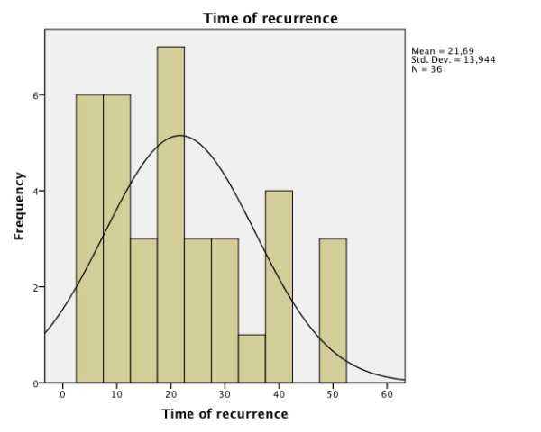
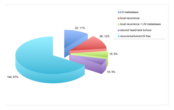
Results: Out of 315 patients with an oral squamous cell carcinoma, 294 were evaluated. Those experiencing neither recurrence of the tumor nor lymph node metastases constituted 62%. Local recurrence was seen in 36 (12%), lymph node metastases in 32 (11%), and both in 16 (6%). Of the 32 patients with lymph node metastases, 25 were recognized first clinically, and 7 were detected by routine CT scans; concerning local recurrence, 32 appeared clinically, and 4 were detected by routine CT scans.
Conclusion: Routine CT for follow-up is still indicated for detecting lymph node metastases as well as local recurrence.


 分享
分享

 求助内容:
求助内容: 应助结果提醒方式:
 扫码关注我们
