{"title":"HOXA4蛋白在主动脉和人腹主动脉瘤中的水平和定位。","authors":"Christian Klausen, Nelly Auersperg","doi":"10.1186/1472-6793-11-18","DOIUrl":null,"url":null,"abstract":"<p><p>This report presents evidence for the specificities of select commercially available HOXA4 antibodies in regards to concerns about the specificity of the HOXA4 antibody used by Lillvis et al. (Regional expression of HOXA4 along the aorta and its potential role in human abdominal aortic aneurysms. BMC Physiol 2011, 11:9). Using an antibody characterized extensively by us, Lillvis et al. report detecting HOXA4 at a size of 33 kDa despite our previous reports that HOXA4 is detected at ~37-39 kDa and that the ~30-33 kDa band is non-specific. Using small interfering RNA targeting HOXA4, forced expression of full-length HOXA4 and HOXA4-positive and -negative ovarian cancer cell lines, we confirm our previous findings that the ~30-33 kDa band is non-specific and that HOXA4 is detected at ~37-39 kDa. Moreover, we demonstrate that HOXA4 small interfering RNA reduces the ~37-39 kDa HOXA4 band, but not the ~30-33 kDa non-specific band, in a human acute monocytic leukemia cell line used by Lillvis et al. Western blot analysis performed with two additional commercially available HOXA4 antibodies also detected HOXA4 at ~37-39 kDa. Lastly, immunofluorescent staining of a HOXA4-negative ovarian cancer cell line with the antibody used by Lillvis et al. yields strong perinuclear staining, similar to that observed by Lillvis et al., which cannot be attributed to HOXA4. Our results highlight and briefly discuss the importance of careful antibody validation and selection for use in various applications.</p>","PeriodicalId":35905,"journal":{"name":"BMC Physiology","volume":"11 ","pages":"18"},"PeriodicalIF":0.0000,"publicationDate":"2011-12-14","publicationTypes":"Journal Article","fieldsOfStudy":null,"isOpenAccess":false,"openAccessPdf":"https://sci-hub-pdf.com/10.1186/1472-6793-11-18","citationCount":"0","resultStr":"{\"title\":\"HOXA4 protein levels and localization in the aorta and in human abdominal aortic aneurysms.\",\"authors\":\"Christian Klausen, Nelly Auersperg\",\"doi\":\"10.1186/1472-6793-11-18\",\"DOIUrl\":null,\"url\":null,\"abstract\":\"<p><p>This report presents evidence for the specificities of select commercially available HOXA4 antibodies in regards to concerns about the specificity of the HOXA4 antibody used by Lillvis et al. (Regional expression of HOXA4 along the aorta and its potential role in human abdominal aortic aneurysms. BMC Physiol 2011, 11:9). Using an antibody characterized extensively by us, Lillvis et al. report detecting HOXA4 at a size of 33 kDa despite our previous reports that HOXA4 is detected at ~37-39 kDa and that the ~30-33 kDa band is non-specific. Using small interfering RNA targeting HOXA4, forced expression of full-length HOXA4 and HOXA4-positive and -negative ovarian cancer cell lines, we confirm our previous findings that the ~30-33 kDa band is non-specific and that HOXA4 is detected at ~37-39 kDa. Moreover, we demonstrate that HOXA4 small interfering RNA reduces the ~37-39 kDa HOXA4 band, but not the ~30-33 kDa non-specific band, in a human acute monocytic leukemia cell line used by Lillvis et al. Western blot analysis performed with two additional commercially available HOXA4 antibodies also detected HOXA4 at ~37-39 kDa. Lastly, immunofluorescent staining of a HOXA4-negative ovarian cancer cell line with the antibody used by Lillvis et al. yields strong perinuclear staining, similar to that observed by Lillvis et al., which cannot be attributed to HOXA4. Our results highlight and briefly discuss the importance of careful antibody validation and selection for use in various applications.</p>\",\"PeriodicalId\":35905,\"journal\":{\"name\":\"BMC Physiology\",\"volume\":\"11 \",\"pages\":\"18\"},\"PeriodicalIF\":0.0000,\"publicationDate\":\"2011-12-14\",\"publicationTypes\":\"Journal Article\",\"fieldsOfStudy\":null,\"isOpenAccess\":false,\"openAccessPdf\":\"https://sci-hub-pdf.com/10.1186/1472-6793-11-18\",\"citationCount\":\"0\",\"resultStr\":null,\"platform\":\"Semanticscholar\",\"paperid\":null,\"PeriodicalName\":\"BMC Physiology\",\"FirstCategoryId\":\"1085\",\"ListUrlMain\":\"https://doi.org/10.1186/1472-6793-11-18\",\"RegionNum\":0,\"RegionCategory\":null,\"ArticlePicture\":[],\"TitleCN\":null,\"AbstractTextCN\":null,\"PMCID\":null,\"EPubDate\":\"\",\"PubModel\":\"\",\"JCR\":\"Q1\",\"JCRName\":\"Biochemistry, Genetics and Molecular Biology\",\"Score\":null,\"Total\":0}","platform":"Semanticscholar","paperid":null,"PeriodicalName":"BMC Physiology","FirstCategoryId":"1085","ListUrlMain":"https://doi.org/10.1186/1472-6793-11-18","RegionNum":0,"RegionCategory":null,"ArticlePicture":[],"TitleCN":null,"AbstractTextCN":null,"PMCID":null,"EPubDate":"","PubModel":"","JCR":"Q1","JCRName":"Biochemistry, Genetics and Molecular Biology","Score":null,"Total":0}
HOXA4 protein levels and localization in the aorta and in human abdominal aortic aneurysms.
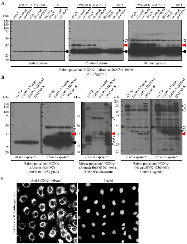
This report presents evidence for the specificities of select commercially available HOXA4 antibodies in regards to concerns about the specificity of the HOXA4 antibody used by Lillvis et al. (Regional expression of HOXA4 along the aorta and its potential role in human abdominal aortic aneurysms. BMC Physiol 2011, 11:9). Using an antibody characterized extensively by us, Lillvis et al. report detecting HOXA4 at a size of 33 kDa despite our previous reports that HOXA4 is detected at ~37-39 kDa and that the ~30-33 kDa band is non-specific. Using small interfering RNA targeting HOXA4, forced expression of full-length HOXA4 and HOXA4-positive and -negative ovarian cancer cell lines, we confirm our previous findings that the ~30-33 kDa band is non-specific and that HOXA4 is detected at ~37-39 kDa. Moreover, we demonstrate that HOXA4 small interfering RNA reduces the ~37-39 kDa HOXA4 band, but not the ~30-33 kDa non-specific band, in a human acute monocytic leukemia cell line used by Lillvis et al. Western blot analysis performed with two additional commercially available HOXA4 antibodies also detected HOXA4 at ~37-39 kDa. Lastly, immunofluorescent staining of a HOXA4-negative ovarian cancer cell line with the antibody used by Lillvis et al. yields strong perinuclear staining, similar to that observed by Lillvis et al., which cannot be attributed to HOXA4. Our results highlight and briefly discuss the importance of careful antibody validation and selection for use in various applications.
BMC PhysiologyBiochemistry, Genetics and Molecular Biology-Physiology
CiteScore
9.60
自引率
0.00%
发文量
0
期刊介绍:
BMC Physiology is an open access journal publishing original peer-reviewed research articles in cellular, tissue-level, organismal, functional, and developmental aspects of physiological processes. BMC Physiology (ISSN 1472-6793) is indexed/tracked/covered by PubMed, MEDLINE, BIOSIS, CAS, EMBASE, Scopus, Zoological Record and Google Scholar.


 分享
分享
 求助内容:
求助内容: 应助结果提醒方式:
 扫码关注我们
