{"title":"肝粘液囊腺瘤伴卵巢样间质:完全切除的必要性。","authors":"Myung Hee Yoon, Ju Won Yoon, Byung Hoon Han","doi":"10.4174/jkss.2011.81.Suppl1.S51","DOIUrl":null,"url":null,"abstract":"<p><p>Cystadenoma of the liver is a rare neoplasm. Although many cystadenomas are asymptomatic, symptoms can include abdominal pain, postprandial epigastric discomfort, and nausea. Dramatic changes in hepatic imaging techniques have been helpful for diagnosing cystic lesions of the liver, such as simple cyst, hydatid cyst, cystadenoma, cystadenocarcinoma, and metastatic neuroendocrine tumors. However, it remains difficult to differentiate cystadenoma from cystadenocarcinoma for multiseptated cystic hepatic lesions with papillary projection on computed tomography (CT) and magnetic resonance imaging (MRI). Here we report the case of a 47-year-old woman with several months of postprandial discomfort and abdominal fullness. CT and MRI revealed multiseptated cystic lesions with papillary excrescences. A left hemihepatectomy was performed. Histology showed a benign mucinous cystic tumor with ovarian-like stroma.</p>","PeriodicalId":49157,"journal":{"name":"Journal of the Korean Surgical Society","volume":"81 Suppl 1 ","pages":"S51-4"},"PeriodicalIF":0.0000,"publicationDate":"2011-12-01","publicationTypes":"Journal Article","fieldsOfStudy":null,"isOpenAccess":false,"openAccessPdf":"https://sci-hub-pdf.com/10.4174/jkss.2011.81.Suppl1.S51","citationCount":"4","resultStr":"{\"title\":\"Mucinous cystadenoma of the liver with ovarian-like stroma: the need for complete resection.\",\"authors\":\"Myung Hee Yoon, Ju Won Yoon, Byung Hoon Han\",\"doi\":\"10.4174/jkss.2011.81.Suppl1.S51\",\"DOIUrl\":null,\"url\":null,\"abstract\":\"<p><p>Cystadenoma of the liver is a rare neoplasm. Although many cystadenomas are asymptomatic, symptoms can include abdominal pain, postprandial epigastric discomfort, and nausea. Dramatic changes in hepatic imaging techniques have been helpful for diagnosing cystic lesions of the liver, such as simple cyst, hydatid cyst, cystadenoma, cystadenocarcinoma, and metastatic neuroendocrine tumors. However, it remains difficult to differentiate cystadenoma from cystadenocarcinoma for multiseptated cystic hepatic lesions with papillary projection on computed tomography (CT) and magnetic resonance imaging (MRI). Here we report the case of a 47-year-old woman with several months of postprandial discomfort and abdominal fullness. CT and MRI revealed multiseptated cystic lesions with papillary excrescences. A left hemihepatectomy was performed. Histology showed a benign mucinous cystic tumor with ovarian-like stroma.</p>\",\"PeriodicalId\":49157,\"journal\":{\"name\":\"Journal of the Korean Surgical Society\",\"volume\":\"81 Suppl 1 \",\"pages\":\"S51-4\"},\"PeriodicalIF\":0.0000,\"publicationDate\":\"2011-12-01\",\"publicationTypes\":\"Journal Article\",\"fieldsOfStudy\":null,\"isOpenAccess\":false,\"openAccessPdf\":\"https://sci-hub-pdf.com/10.4174/jkss.2011.81.Suppl1.S51\",\"citationCount\":\"4\",\"resultStr\":null,\"platform\":\"Semanticscholar\",\"paperid\":null,\"PeriodicalName\":\"Journal of the Korean Surgical Society\",\"FirstCategoryId\":\"1085\",\"ListUrlMain\":\"https://doi.org/10.4174/jkss.2011.81.Suppl1.S51\",\"RegionNum\":0,\"RegionCategory\":null,\"ArticlePicture\":[],\"TitleCN\":null,\"AbstractTextCN\":null,\"PMCID\":null,\"EPubDate\":\"2011/11/25 0:00:00\",\"PubModel\":\"Epub\",\"JCR\":\"\",\"JCRName\":\"\",\"Score\":null,\"Total\":0}","platform":"Semanticscholar","paperid":null,"PeriodicalName":"Journal of the Korean Surgical Society","FirstCategoryId":"1085","ListUrlMain":"https://doi.org/10.4174/jkss.2011.81.Suppl1.S51","RegionNum":0,"RegionCategory":null,"ArticlePicture":[],"TitleCN":null,"AbstractTextCN":null,"PMCID":null,"EPubDate":"2011/11/25 0:00:00","PubModel":"Epub","JCR":"","JCRName":"","Score":null,"Total":0}
Mucinous cystadenoma of the liver with ovarian-like stroma: the need for complete resection.
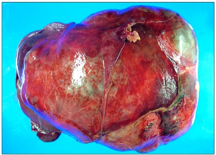
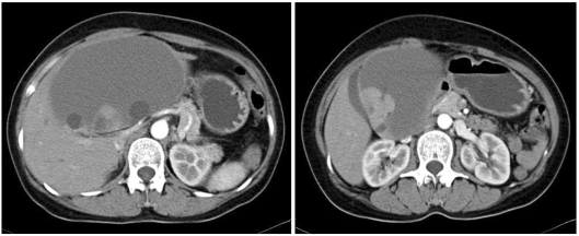
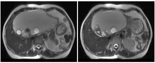
Cystadenoma of the liver is a rare neoplasm. Although many cystadenomas are asymptomatic, symptoms can include abdominal pain, postprandial epigastric discomfort, and nausea. Dramatic changes in hepatic imaging techniques have been helpful for diagnosing cystic lesions of the liver, such as simple cyst, hydatid cyst, cystadenoma, cystadenocarcinoma, and metastatic neuroendocrine tumors. However, it remains difficult to differentiate cystadenoma from cystadenocarcinoma for multiseptated cystic hepatic lesions with papillary projection on computed tomography (CT) and magnetic resonance imaging (MRI). Here we report the case of a 47-year-old woman with several months of postprandial discomfort and abdominal fullness. CT and MRI revealed multiseptated cystic lesions with papillary excrescences. A left hemihepatectomy was performed. Histology showed a benign mucinous cystic tumor with ovarian-like stroma.


 分享
分享
 求助内容:
求助内容: 应助结果提醒方式:
 扫码关注我们
