Eun Young Kim, In Kyu Lee, Yoon Suk Lee, Naery Yang, Dong Jin Chung, Kwang-Il Yim, Jin Il Kim, Seung Taek Oh
{"title":"结肠炎性肌成纤维细胞瘤。","authors":"Eun Young Kim, In Kyu Lee, Yoon Suk Lee, Naery Yang, Dong Jin Chung, Kwang-Il Yim, Jin Il Kim, Seung Taek Oh","doi":"10.4174/jkss.2012.82.1.45","DOIUrl":null,"url":null,"abstract":"<p><p>Inflammatory myofibroblastic tumor (IMT) is an uncommon mesenchymal solid tumor commonly documented in children and young adults. Here, we report a case of IMT in colon confirmed pathologically after laparoscopic anterior resection. A 35-year-old man presented with anal bleeding after defecation for 2 weeks. Colonoscopy demonstrated a mass with shallow ulceration in the central area and irregular margin accompanied by intact mucosa in the descending colon. Computer tomography showed a well-demarcated and homogenous solitary mass in the descending colon. We performed laparoscopic anterior resection. This case was diagnosed as IMT after microscopic examination. The tumor was composed of a proliferation of spindle-shaped cells arranged in the hyaline material with chronic inflammatory cells, composed mainly of plasma cells and lymphocytes. Immunohistochemically, tumor cells were positive for smooth muscle actin, and vimentin, and negative for desmin, CD117 (c-kit), anaplastic lymphoma kinase-1.</p>","PeriodicalId":49157,"journal":{"name":"Journal of the Korean Surgical Society","volume":"82 1","pages":"45-9"},"PeriodicalIF":0.0000,"publicationDate":"2012-01-01","publicationTypes":"Journal Article","fieldsOfStudy":null,"isOpenAccess":false,"openAccessPdf":"https://sci-hub-pdf.com/10.4174/jkss.2012.82.1.45","citationCount":"47","resultStr":"{\"title\":\"Inflammatory myofibroblastic tumor in colon.\",\"authors\":\"Eun Young Kim, In Kyu Lee, Yoon Suk Lee, Naery Yang, Dong Jin Chung, Kwang-Il Yim, Jin Il Kim, Seung Taek Oh\",\"doi\":\"10.4174/jkss.2012.82.1.45\",\"DOIUrl\":null,\"url\":null,\"abstract\":\"<p><p>Inflammatory myofibroblastic tumor (IMT) is an uncommon mesenchymal solid tumor commonly documented in children and young adults. Here, we report a case of IMT in colon confirmed pathologically after laparoscopic anterior resection. A 35-year-old man presented with anal bleeding after defecation for 2 weeks. Colonoscopy demonstrated a mass with shallow ulceration in the central area and irregular margin accompanied by intact mucosa in the descending colon. Computer tomography showed a well-demarcated and homogenous solitary mass in the descending colon. We performed laparoscopic anterior resection. This case was diagnosed as IMT after microscopic examination. The tumor was composed of a proliferation of spindle-shaped cells arranged in the hyaline material with chronic inflammatory cells, composed mainly of plasma cells and lymphocytes. Immunohistochemically, tumor cells were positive for smooth muscle actin, and vimentin, and negative for desmin, CD117 (c-kit), anaplastic lymphoma kinase-1.</p>\",\"PeriodicalId\":49157,\"journal\":{\"name\":\"Journal of the Korean Surgical Society\",\"volume\":\"82 1\",\"pages\":\"45-9\"},\"PeriodicalIF\":0.0000,\"publicationDate\":\"2012-01-01\",\"publicationTypes\":\"Journal Article\",\"fieldsOfStudy\":null,\"isOpenAccess\":false,\"openAccessPdf\":\"https://sci-hub-pdf.com/10.4174/jkss.2012.82.1.45\",\"citationCount\":\"47\",\"resultStr\":null,\"platform\":\"Semanticscholar\",\"paperid\":null,\"PeriodicalName\":\"Journal of the Korean Surgical Society\",\"FirstCategoryId\":\"1085\",\"ListUrlMain\":\"https://doi.org/10.4174/jkss.2012.82.1.45\",\"RegionNum\":0,\"RegionCategory\":null,\"ArticlePicture\":[],\"TitleCN\":null,\"AbstractTextCN\":null,\"PMCID\":null,\"EPubDate\":\"2011/12/27 0:00:00\",\"PubModel\":\"Epub\",\"JCR\":\"\",\"JCRName\":\"\",\"Score\":null,\"Total\":0}","platform":"Semanticscholar","paperid":null,"PeriodicalName":"Journal of the Korean Surgical Society","FirstCategoryId":"1085","ListUrlMain":"https://doi.org/10.4174/jkss.2012.82.1.45","RegionNum":0,"RegionCategory":null,"ArticlePicture":[],"TitleCN":null,"AbstractTextCN":null,"PMCID":null,"EPubDate":"2011/12/27 0:00:00","PubModel":"Epub","JCR":"","JCRName":"","Score":null,"Total":0}
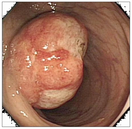
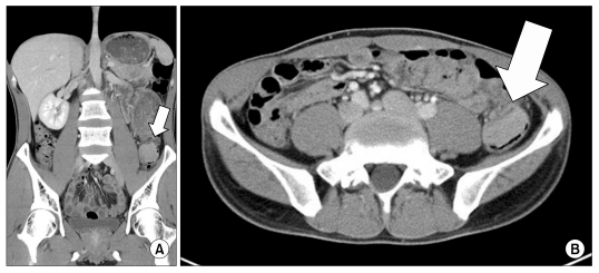
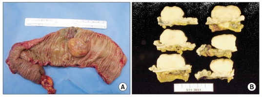
Inflammatory myofibroblastic tumor (IMT) is an uncommon mesenchymal solid tumor commonly documented in children and young adults. Here, we report a case of IMT in colon confirmed pathologically after laparoscopic anterior resection. A 35-year-old man presented with anal bleeding after defecation for 2 weeks. Colonoscopy demonstrated a mass with shallow ulceration in the central area and irregular margin accompanied by intact mucosa in the descending colon. Computer tomography showed a well-demarcated and homogenous solitary mass in the descending colon. We performed laparoscopic anterior resection. This case was diagnosed as IMT after microscopic examination. The tumor was composed of a proliferation of spindle-shaped cells arranged in the hyaline material with chronic inflammatory cells, composed mainly of plasma cells and lymphocytes. Immunohistochemically, tumor cells were positive for smooth muscle actin, and vimentin, and negative for desmin, CD117 (c-kit), anaplastic lymphoma kinase-1.


 分享
分享
 求助内容:
求助内容: 应助结果提醒方式:
 扫码关注我们
