{"title":"2例1型和2型x连锁淋巴细胞增生性综合征患者EBV相关噬血细胞性淋巴组织细胞增多症中EBV感染细胞的特征","authors":"Xi Yang, Taizo Wada, Ken-Ichi Imadome, Naonori Nishida, Takeo Mukai, Mitsuhiro Fujiwara, Haruka Kawashima, Fumiyo Kato, Shigeyoshi Fujiwara, Akihiro Yachie, Xiaodong Zhao, Toshio Miyawaki, Hirokazu Kanegane","doi":"10.1186/2042-4280-3-1","DOIUrl":null,"url":null,"abstract":"<p><strong>Background: </strong>X-linked lymphoproliferative syndrome (XLP) is a rare inherited immunodeficiency by an extreme vulnerability to Epstein-Barr virus (EBV) infection, frequently resulting in hemophagocytic lymphohistiocytosis (HLH). XLP are now divided into type 1 (XLP-1) and type 2 (XLP-2), which are caused by mutations of SH2D1A/SLAM-associated protein (SAP) and X-linked inhibitor of apoptosis protein (XIAP) genes, respectively. The diagnosis of XLP in individuals with EBV-associated HLH (EBV-HLH) is generally difficult because they show basically similar symptoms to sporadic EBV-HLH. Although EBV-infected cells in sporadic EBV-HLH are known to be mainly in CD8+ T cells, the cell-type of EBV-infected cells in EBV-HLH seen in XLP patients remains undetermined.</p><p><strong>Methods: </strong>EBV-infected cells in two patients (XLP-1 and XLP-2) presenting EBV-HLH were evaluated by in EBER-1 in situ hybridization or quantitative PCR methods.</p><p><strong>Results: </strong>Both XLP patients showed that the dominant population of EBV-infected cells was CD19+ B cells, whereas EBV-infected CD8+ T cells were very few.</p><p><strong>Conclusions: </strong>In XLP-related EBV-HLH, EBV-infected cells appear to be predominantly B cells. B cell directed therapy such as rituximab may be a valuable option in the treatment of EBV-HLH in XLP patients.</p>","PeriodicalId":89143,"journal":{"name":"Herpesviridae","volume":"3 1","pages":"1"},"PeriodicalIF":0.0000,"publicationDate":"2012-02-10","publicationTypes":"Journal Article","fieldsOfStudy":null,"isOpenAccess":false,"openAccessPdf":"https://sci-hub-pdf.com/10.1186/2042-4280-3-1","citationCount":"23","resultStr":"{\"title\":\"Characterization of Epstein-Barr virus (EBV)-infected cells in EBV-associated hemophagocytic lymphohistiocytosis in two patients with X-linked lymphoproliferative syndrome type 1 and type 2.\",\"authors\":\"Xi Yang, Taizo Wada, Ken-Ichi Imadome, Naonori Nishida, Takeo Mukai, Mitsuhiro Fujiwara, Haruka Kawashima, Fumiyo Kato, Shigeyoshi Fujiwara, Akihiro Yachie, Xiaodong Zhao, Toshio Miyawaki, Hirokazu Kanegane\",\"doi\":\"10.1186/2042-4280-3-1\",\"DOIUrl\":null,\"url\":null,\"abstract\":\"<p><strong>Background: </strong>X-linked lymphoproliferative syndrome (XLP) is a rare inherited immunodeficiency by an extreme vulnerability to Epstein-Barr virus (EBV) infection, frequently resulting in hemophagocytic lymphohistiocytosis (HLH). XLP are now divided into type 1 (XLP-1) and type 2 (XLP-2), which are caused by mutations of SH2D1A/SLAM-associated protein (SAP) and X-linked inhibitor of apoptosis protein (XIAP) genes, respectively. The diagnosis of XLP in individuals with EBV-associated HLH (EBV-HLH) is generally difficult because they show basically similar symptoms to sporadic EBV-HLH. Although EBV-infected cells in sporadic EBV-HLH are known to be mainly in CD8+ T cells, the cell-type of EBV-infected cells in EBV-HLH seen in XLP patients remains undetermined.</p><p><strong>Methods: </strong>EBV-infected cells in two patients (XLP-1 and XLP-2) presenting EBV-HLH were evaluated by in EBER-1 in situ hybridization or quantitative PCR methods.</p><p><strong>Results: </strong>Both XLP patients showed that the dominant population of EBV-infected cells was CD19+ B cells, whereas EBV-infected CD8+ T cells were very few.</p><p><strong>Conclusions: </strong>In XLP-related EBV-HLH, EBV-infected cells appear to be predominantly B cells. B cell directed therapy such as rituximab may be a valuable option in the treatment of EBV-HLH in XLP patients.</p>\",\"PeriodicalId\":89143,\"journal\":{\"name\":\"Herpesviridae\",\"volume\":\"3 1\",\"pages\":\"1\"},\"PeriodicalIF\":0.0000,\"publicationDate\":\"2012-02-10\",\"publicationTypes\":\"Journal Article\",\"fieldsOfStudy\":null,\"isOpenAccess\":false,\"openAccessPdf\":\"https://sci-hub-pdf.com/10.1186/2042-4280-3-1\",\"citationCount\":\"23\",\"resultStr\":null,\"platform\":\"Semanticscholar\",\"paperid\":null,\"PeriodicalName\":\"Herpesviridae\",\"FirstCategoryId\":\"1085\",\"ListUrlMain\":\"https://doi.org/10.1186/2042-4280-3-1\",\"RegionNum\":0,\"RegionCategory\":null,\"ArticlePicture\":[],\"TitleCN\":null,\"AbstractTextCN\":null,\"PMCID\":null,\"EPubDate\":\"\",\"PubModel\":\"\",\"JCR\":\"\",\"JCRName\":\"\",\"Score\":null,\"Total\":0}","platform":"Semanticscholar","paperid":null,"PeriodicalName":"Herpesviridae","FirstCategoryId":"1085","ListUrlMain":"https://doi.org/10.1186/2042-4280-3-1","RegionNum":0,"RegionCategory":null,"ArticlePicture":[],"TitleCN":null,"AbstractTextCN":null,"PMCID":null,"EPubDate":"","PubModel":"","JCR":"","JCRName":"","Score":null,"Total":0}
引用次数: 23
摘要
背景:x连锁淋巴细胞增生性综合征(XLP)是一种罕见的遗传性免疫缺陷,由eb病毒(EBV)感染的极端易感性引起,经常导致噬血细胞性淋巴组织细胞增多症(HLH)。XLP目前分为1型(XLP-1)和2型(XLP-2),分别由SH2D1A/SLAM-associated protein (SAP)和X-linked inhibitor of apoptosis protein (XIAP)基因突变引起。ebv相关HLH (EBV-HLH)患者的XLP诊断通常很困难,因为他们表现出与散发的EBV-HLH基本相似的症状。虽然已知散发性EBV-HLH中的ebv感染细胞主要是CD8+ T细胞,但XLP患者中EBV-HLH中ebv感染细胞的细胞类型仍不确定。方法:对2例EBV-HLH患者(XLP-1和XLP-2)的ebv感染细胞进行原位杂交或定量PCR检测。结果:两例XLP患者ebv感染细胞均以CD19+ B细胞为主,而感染的CD8+ T细胞极少。结论:在xlp相关的EBV-HLH中,ebv感染的细胞似乎主要是B细胞。B细胞定向治疗如利妥昔单抗可能是治疗XLP患者EBV-HLH的一个有价值的选择。
Characterization of Epstein-Barr virus (EBV)-infected cells in EBV-associated hemophagocytic lymphohistiocytosis in two patients with X-linked lymphoproliferative syndrome type 1 and type 2.
Background: X-linked lymphoproliferative syndrome (XLP) is a rare inherited immunodeficiency by an extreme vulnerability to Epstein-Barr virus (EBV) infection, frequently resulting in hemophagocytic lymphohistiocytosis (HLH). XLP are now divided into type 1 (XLP-1) and type 2 (XLP-2), which are caused by mutations of SH2D1A/SLAM-associated protein (SAP) and X-linked inhibitor of apoptosis protein (XIAP) genes, respectively. The diagnosis of XLP in individuals with EBV-associated HLH (EBV-HLH) is generally difficult because they show basically similar symptoms to sporadic EBV-HLH. Although EBV-infected cells in sporadic EBV-HLH are known to be mainly in CD8+ T cells, the cell-type of EBV-infected cells in EBV-HLH seen in XLP patients remains undetermined.
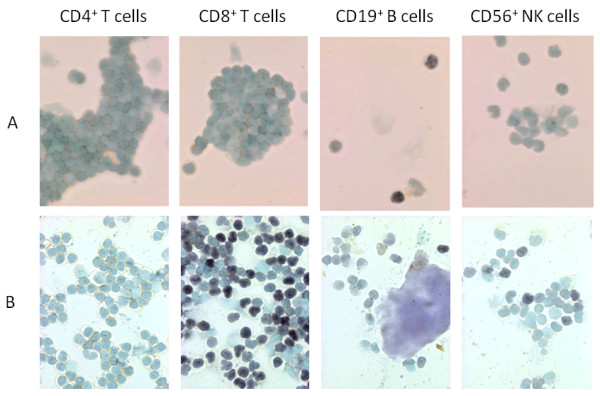
Methods: EBV-infected cells in two patients (XLP-1 and XLP-2) presenting EBV-HLH were evaluated by in EBER-1 in situ hybridization or quantitative PCR methods.
Results: Both XLP patients showed that the dominant population of EBV-infected cells was CD19+ B cells, whereas EBV-infected CD8+ T cells were very few.
Conclusions: In XLP-related EBV-HLH, EBV-infected cells appear to be predominantly B cells. B cell directed therapy such as rituximab may be a valuable option in the treatment of EBV-HLH in XLP patients.


 分享
分享

 求助内容:
求助内容: 应助结果提醒方式:
 扫码关注我们
