{"title":"胫骨应力损伤的高分辨率轴向磁共振成像。","authors":"Takeo Mammoto, Atsushi Hirano, Yohei Tomaru, Mamoru Kono, Yuta Tsukagoshi, Sinzo Onishi, Naotaka Mamizuka","doi":"10.1186/1758-2555-4-16","DOIUrl":null,"url":null,"abstract":"<p><strong>Purpose: </strong>To evaluate the relative involvement of tibial stress injuries using high-resolution axial MR imaging and the correlation with MR and radiographic images.</p><p><strong>Methods: </strong>A total of 33 patients with exercise-induced tibial pain were evaluated. All patients underwent radiograph and high-resolution axial MR imaging. Radiographs were taken at initial presentation and 4 weeks later. High-resolution MR axial images were obtained using a microscopy surface coil with 60 × 60 mm field of view on a 1.5T MR unit. All images were evaluated for abnormal signals of the periosteum, cortex and bone marrow.</p><p><strong>Results: </strong>Nineteen patients showed no periosteal reaction at initial and follow-up radiographs. MR imaging showed abnormal signals in the periosteal tissue and partially abnormal signals in the bone marrow. In 7 patients, periosteal reaction was not seen at initial radiograph, but was detected at follow-up radiograph. MR imaging showed abnormal signals in the periosteal tissue and entire bone marrow. Abnormal signals in the cortex were found in 6 patients. The remaining 7 showed periosteal reactions at initial radiograph. MR imaging showed abnormal signals in the periosteal tissue in 6 patients. Abnormal signals were seen in the partial and entire bone marrow in 4 and 3 patients, respectively.</p><p><strong>Conclusions: </strong>Bone marrow abnormalities in high-resolution axial MR imaging were related to periosteal reactions at follow-up radiograph. Bone marrow abnormalities might predict later periosteal reactions, suggesting shin splints or stress fractures. High-resolution axial MR imaging is useful in early discrimination of tibial stress injuries.</p>","PeriodicalId":88316,"journal":{"name":"Sports medicine, arthroscopy, rehabilitation, therapy & technology : SMARTT","volume":"4 1","pages":"16"},"PeriodicalIF":0.0000,"publicationDate":"2012-05-10","publicationTypes":"Journal Article","fieldsOfStudy":null,"isOpenAccess":false,"openAccessPdf":"https://sci-hub-pdf.com/10.1186/1758-2555-4-16","citationCount":"8","resultStr":"{\"title\":\"High-resolution axial MR imaging of tibial stress injuries.\",\"authors\":\"Takeo Mammoto, Atsushi Hirano, Yohei Tomaru, Mamoru Kono, Yuta Tsukagoshi, Sinzo Onishi, Naotaka Mamizuka\",\"doi\":\"10.1186/1758-2555-4-16\",\"DOIUrl\":null,\"url\":null,\"abstract\":\"<p><strong>Purpose: </strong>To evaluate the relative involvement of tibial stress injuries using high-resolution axial MR imaging and the correlation with MR and radiographic images.</p><p><strong>Methods: </strong>A total of 33 patients with exercise-induced tibial pain were evaluated. All patients underwent radiograph and high-resolution axial MR imaging. Radiographs were taken at initial presentation and 4 weeks later. High-resolution MR axial images were obtained using a microscopy surface coil with 60 × 60 mm field of view on a 1.5T MR unit. All images were evaluated for abnormal signals of the periosteum, cortex and bone marrow.</p><p><strong>Results: </strong>Nineteen patients showed no periosteal reaction at initial and follow-up radiographs. MR imaging showed abnormal signals in the periosteal tissue and partially abnormal signals in the bone marrow. In 7 patients, periosteal reaction was not seen at initial radiograph, but was detected at follow-up radiograph. MR imaging showed abnormal signals in the periosteal tissue and entire bone marrow. Abnormal signals in the cortex were found in 6 patients. The remaining 7 showed periosteal reactions at initial radiograph. MR imaging showed abnormal signals in the periosteal tissue in 6 patients. Abnormal signals were seen in the partial and entire bone marrow in 4 and 3 patients, respectively.</p><p><strong>Conclusions: </strong>Bone marrow abnormalities in high-resolution axial MR imaging were related to periosteal reactions at follow-up radiograph. Bone marrow abnormalities might predict later periosteal reactions, suggesting shin splints or stress fractures. High-resolution axial MR imaging is useful in early discrimination of tibial stress injuries.</p>\",\"PeriodicalId\":88316,\"journal\":{\"name\":\"Sports medicine, arthroscopy, rehabilitation, therapy & technology : SMARTT\",\"volume\":\"4 1\",\"pages\":\"16\"},\"PeriodicalIF\":0.0000,\"publicationDate\":\"2012-05-10\",\"publicationTypes\":\"Journal Article\",\"fieldsOfStudy\":null,\"isOpenAccess\":false,\"openAccessPdf\":\"https://sci-hub-pdf.com/10.1186/1758-2555-4-16\",\"citationCount\":\"8\",\"resultStr\":null,\"platform\":\"Semanticscholar\",\"paperid\":null,\"PeriodicalName\":\"Sports medicine, arthroscopy, rehabilitation, therapy & technology : SMARTT\",\"FirstCategoryId\":\"1085\",\"ListUrlMain\":\"https://doi.org/10.1186/1758-2555-4-16\",\"RegionNum\":0,\"RegionCategory\":null,\"ArticlePicture\":[],\"TitleCN\":null,\"AbstractTextCN\":null,\"PMCID\":null,\"EPubDate\":\"\",\"PubModel\":\"\",\"JCR\":\"\",\"JCRName\":\"\",\"Score\":null,\"Total\":0}","platform":"Semanticscholar","paperid":null,"PeriodicalName":"Sports medicine, arthroscopy, rehabilitation, therapy & technology : SMARTT","FirstCategoryId":"1085","ListUrlMain":"https://doi.org/10.1186/1758-2555-4-16","RegionNum":0,"RegionCategory":null,"ArticlePicture":[],"TitleCN":null,"AbstractTextCN":null,"PMCID":null,"EPubDate":"","PubModel":"","JCR":"","JCRName":"","Score":null,"Total":0}
High-resolution axial MR imaging of tibial stress injuries.
Purpose: To evaluate the relative involvement of tibial stress injuries using high-resolution axial MR imaging and the correlation with MR and radiographic images.
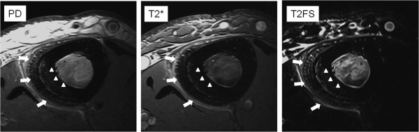
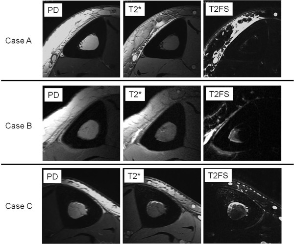
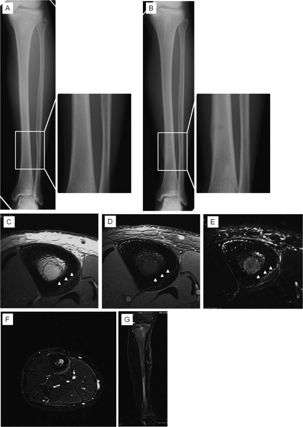
Methods: A total of 33 patients with exercise-induced tibial pain were evaluated. All patients underwent radiograph and high-resolution axial MR imaging. Radiographs were taken at initial presentation and 4 weeks later. High-resolution MR axial images were obtained using a microscopy surface coil with 60 × 60 mm field of view on a 1.5T MR unit. All images were evaluated for abnormal signals of the periosteum, cortex and bone marrow.
Results: Nineteen patients showed no periosteal reaction at initial and follow-up radiographs. MR imaging showed abnormal signals in the periosteal tissue and partially abnormal signals in the bone marrow. In 7 patients, periosteal reaction was not seen at initial radiograph, but was detected at follow-up radiograph. MR imaging showed abnormal signals in the periosteal tissue and entire bone marrow. Abnormal signals in the cortex were found in 6 patients. The remaining 7 showed periosteal reactions at initial radiograph. MR imaging showed abnormal signals in the periosteal tissue in 6 patients. Abnormal signals were seen in the partial and entire bone marrow in 4 and 3 patients, respectively.
Conclusions: Bone marrow abnormalities in high-resolution axial MR imaging were related to periosteal reactions at follow-up radiograph. Bone marrow abnormalities might predict later periosteal reactions, suggesting shin splints or stress fractures. High-resolution axial MR imaging is useful in early discrimination of tibial stress injuries.


 分享
分享

 求助内容:
求助内容: 应助结果提醒方式:
 扫码关注我们
