Zaid Hamdoon, Waseem Jerjes, Raed Al-Delayme, Gordon McKenzie, Amrita Jay, Colin Hopper
{"title":"光学相干断层扫描口腔粘膜组织的结构验证。","authors":"Zaid Hamdoon, Waseem Jerjes, Raed Al-Delayme, Gordon McKenzie, Amrita Jay, Colin Hopper","doi":"10.1186/1758-3284-4-29","DOIUrl":null,"url":null,"abstract":"<p><strong>Background: </strong>Optical coherence tomography (OCT) is a non-invasive optical technology using near-infrared light to produce cross-sectional tissue images with lateral resolution.</p><p><strong>Objectives: </strong>The overall aims of this study was to generate a bank of normative and pathological OCT data of the oral tissues to allow identification of cellular structures of normal and pathological processes with the aim to create a diagnostic algorithm which can be used in the early detection of oral disorders.</p><p><strong>Material and methods: </strong>Seventy-three patients with 78 suspicious oral lesions were referred for further management to the UCLH Head and Neck Centre, London. The entire cohort had their lesions surgically biopsied (incisional or excisional). The immediate ex vivo phase involved scanning the specimens using optical coherence tomography. The specimens were then processed by a histopathologist. Five tissue structures were evaluated as part of this study, including: keratin cell layer, epithelial layer, basement membrane, lamina propria and other microanatomical structures. Two independent assessors (clinician and pathologist trained to use OCT) assessed the OCT images and were asked to comment on the cellular structures and changes involving the five tissue structures in non-blind fashion.</p><p><strong>Results: </strong>Correct identification of the keratin cell layer and its structural changes was achieved in 87% of the cohort; for the epithelial layer it reached 93.5%, and 94% for the basement membrane. Microanatomical structures identification was 64% for blood vessels, 58% for salivary gland ducts and 89% for rete pegs. The agreement was \"good\" between the clinician and the pathologist. OCT was able to differential normal from pathological tissue and pathological tissue of different entities in this immediate ex vivo study. Unfortunately, OCT provided inadequate cellular and subcellular information to enable the grading of oral premalignant disorders.</p><p><strong>Conclusion: </strong>This study enabled the creation of OCT bank of normal and pathological oral tissues. The pathological changes identified using OCT enabled differentiation between normal and pathological tissues, and identification of different tissue pathologies. Further studies are required to assess the accuracy of OCT in identification of various pathological processes involving the oral tissues.</p>","PeriodicalId":49195,"journal":{"name":"Head and Neck Optical Diagnostics Society","volume":"4 ","pages":"29"},"PeriodicalIF":0.0000,"publicationDate":"2012-06-06","publicationTypes":"Journal Article","fieldsOfStudy":null,"isOpenAccess":false,"openAccessPdf":"https://sci-hub-pdf.com/10.1186/1758-3284-4-29","citationCount":"40","resultStr":"{\"title\":\"Structural validation of oral mucosal tissue using optical coherence tomography.\",\"authors\":\"Zaid Hamdoon, Waseem Jerjes, Raed Al-Delayme, Gordon McKenzie, Amrita Jay, Colin Hopper\",\"doi\":\"10.1186/1758-3284-4-29\",\"DOIUrl\":null,\"url\":null,\"abstract\":\"<p><strong>Background: </strong>Optical coherence tomography (OCT) is a non-invasive optical technology using near-infrared light to produce cross-sectional tissue images with lateral resolution.</p><p><strong>Objectives: </strong>The overall aims of this study was to generate a bank of normative and pathological OCT data of the oral tissues to allow identification of cellular structures of normal and pathological processes with the aim to create a diagnostic algorithm which can be used in the early detection of oral disorders.</p><p><strong>Material and methods: </strong>Seventy-three patients with 78 suspicious oral lesions were referred for further management to the UCLH Head and Neck Centre, London. The entire cohort had their lesions surgically biopsied (incisional or excisional). The immediate ex vivo phase involved scanning the specimens using optical coherence tomography. The specimens were then processed by a histopathologist. Five tissue structures were evaluated as part of this study, including: keratin cell layer, epithelial layer, basement membrane, lamina propria and other microanatomical structures. Two independent assessors (clinician and pathologist trained to use OCT) assessed the OCT images and were asked to comment on the cellular structures and changes involving the five tissue structures in non-blind fashion.</p><p><strong>Results: </strong>Correct identification of the keratin cell layer and its structural changes was achieved in 87% of the cohort; for the epithelial layer it reached 93.5%, and 94% for the basement membrane. Microanatomical structures identification was 64% for blood vessels, 58% for salivary gland ducts and 89% for rete pegs. The agreement was \\\"good\\\" between the clinician and the pathologist. OCT was able to differential normal from pathological tissue and pathological tissue of different entities in this immediate ex vivo study. Unfortunately, OCT provided inadequate cellular and subcellular information to enable the grading of oral premalignant disorders.</p><p><strong>Conclusion: </strong>This study enabled the creation of OCT bank of normal and pathological oral tissues. The pathological changes identified using OCT enabled differentiation between normal and pathological tissues, and identification of different tissue pathologies. Further studies are required to assess the accuracy of OCT in identification of various pathological processes involving the oral tissues.</p>\",\"PeriodicalId\":49195,\"journal\":{\"name\":\"Head and Neck Optical Diagnostics Society\",\"volume\":\"4 \",\"pages\":\"29\"},\"PeriodicalIF\":0.0000,\"publicationDate\":\"2012-06-06\",\"publicationTypes\":\"Journal Article\",\"fieldsOfStudy\":null,\"isOpenAccess\":false,\"openAccessPdf\":\"https://sci-hub-pdf.com/10.1186/1758-3284-4-29\",\"citationCount\":\"40\",\"resultStr\":null,\"platform\":\"Semanticscholar\",\"paperid\":null,\"PeriodicalName\":\"Head and Neck Optical Diagnostics Society\",\"FirstCategoryId\":\"1085\",\"ListUrlMain\":\"https://doi.org/10.1186/1758-3284-4-29\",\"RegionNum\":0,\"RegionCategory\":null,\"ArticlePicture\":[],\"TitleCN\":null,\"AbstractTextCN\":null,\"PMCID\":null,\"EPubDate\":\"\",\"PubModel\":\"\",\"JCR\":\"\",\"JCRName\":\"\",\"Score\":null,\"Total\":0}","platform":"Semanticscholar","paperid":null,"PeriodicalName":"Head and Neck Optical Diagnostics Society","FirstCategoryId":"1085","ListUrlMain":"https://doi.org/10.1186/1758-3284-4-29","RegionNum":0,"RegionCategory":null,"ArticlePicture":[],"TitleCN":null,"AbstractTextCN":null,"PMCID":null,"EPubDate":"","PubModel":"","JCR":"","JCRName":"","Score":null,"Total":0}
Structural validation of oral mucosal tissue using optical coherence tomography.
Background: Optical coherence tomography (OCT) is a non-invasive optical technology using near-infrared light to produce cross-sectional tissue images with lateral resolution.
Objectives: The overall aims of this study was to generate a bank of normative and pathological OCT data of the oral tissues to allow identification of cellular structures of normal and pathological processes with the aim to create a diagnostic algorithm which can be used in the early detection of oral disorders.
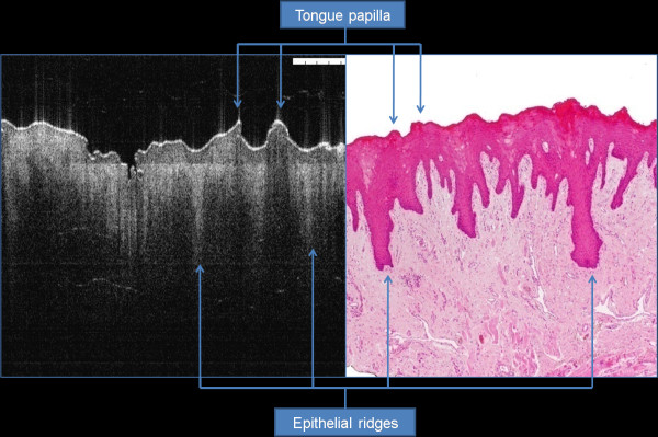
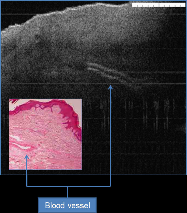
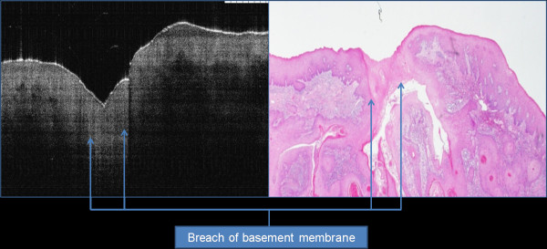
Material and methods: Seventy-three patients with 78 suspicious oral lesions were referred for further management to the UCLH Head and Neck Centre, London. The entire cohort had their lesions surgically biopsied (incisional or excisional). The immediate ex vivo phase involved scanning the specimens using optical coherence tomography. The specimens were then processed by a histopathologist. Five tissue structures were evaluated as part of this study, including: keratin cell layer, epithelial layer, basement membrane, lamina propria and other microanatomical structures. Two independent assessors (clinician and pathologist trained to use OCT) assessed the OCT images and were asked to comment on the cellular structures and changes involving the five tissue structures in non-blind fashion.
Results: Correct identification of the keratin cell layer and its structural changes was achieved in 87% of the cohort; for the epithelial layer it reached 93.5%, and 94% for the basement membrane. Microanatomical structures identification was 64% for blood vessels, 58% for salivary gland ducts and 89% for rete pegs. The agreement was "good" between the clinician and the pathologist. OCT was able to differential normal from pathological tissue and pathological tissue of different entities in this immediate ex vivo study. Unfortunately, OCT provided inadequate cellular and subcellular information to enable the grading of oral premalignant disorders.
Conclusion: This study enabled the creation of OCT bank of normal and pathological oral tissues. The pathological changes identified using OCT enabled differentiation between normal and pathological tissues, and identification of different tissue pathologies. Further studies are required to assess the accuracy of OCT in identification of various pathological processes involving the oral tissues.


 分享
分享
 求助内容:
求助内容: 应助结果提醒方式:
 扫码关注我们
