Molly R Kelly-Goss, Erica R Winterer, Peter C Stapor, Ming Yang, Richard S Sweat, William B Stallcup, Geert W Schmid-Schönbein, Walter L Murfee
{"title":"微血管网生长过程中细胞沿维管岛增殖。","authors":"Molly R Kelly-Goss, Erica R Winterer, Peter C Stapor, Ming Yang, Richard S Sweat, William B Stallcup, Geert W Schmid-Schönbein, Walter L Murfee","doi":"10.1186/1472-6793-12-7","DOIUrl":null,"url":null,"abstract":"<p><strong>Background: </strong>Observations in our laboratory provide evidence of vascular islands, defined as disconnected endothelial cell segments, in the adult microcirculation. The objective of this study was to determine if vascular islands are involved in angiogenesis during microvascular network growth.</p><p><strong>Results: </strong>Mesenteric tissues, which allow visualization of entire microvascular networks at a single cell level, were harvested from unstimulated adult male Wistar rats and Wistar rats 3 and 10 days post angiogenesis stimulation by mast cell degranulation with compound 48/80. Tissues were immunolabeled for PECAM and BRDU. Identification of vessel lumens via injection of FITC-dextran confirmed that endothelial cell segments were disconnected from nearby patent networks. Stimulated networks displayed increases in vascular area, length density, and capillary sprouting. On day 3, the percentage of islands with at least one BRDU-positive cell increased compared to the unstimulated level and was equal to the percentage of capillary sprouts with at least one BRDU-positive cell. At day 10, the number of vascular islands per vascular area dramatically decreased compared to unstimulated and day 3 levels.</p><p><strong>Conclusions: </strong>These results show that vascular islands have the ability to proliferate and suggest that they are able to incorporate into the microcirculation during the initial stages of microvascular network growth.</p>","PeriodicalId":35905,"journal":{"name":"BMC Physiology","volume":"12 ","pages":"7"},"PeriodicalIF":0.0000,"publicationDate":"2012-06-21","publicationTypes":"Journal Article","fieldsOfStudy":null,"isOpenAccess":false,"openAccessPdf":"https://sci-hub-pdf.com/10.1186/1472-6793-12-7","citationCount":"14","resultStr":"{\"title\":\"Cell proliferation along vascular islands during microvascular network growth.\",\"authors\":\"Molly R Kelly-Goss, Erica R Winterer, Peter C Stapor, Ming Yang, Richard S Sweat, William B Stallcup, Geert W Schmid-Schönbein, Walter L Murfee\",\"doi\":\"10.1186/1472-6793-12-7\",\"DOIUrl\":null,\"url\":null,\"abstract\":\"<p><strong>Background: </strong>Observations in our laboratory provide evidence of vascular islands, defined as disconnected endothelial cell segments, in the adult microcirculation. The objective of this study was to determine if vascular islands are involved in angiogenesis during microvascular network growth.</p><p><strong>Results: </strong>Mesenteric tissues, which allow visualization of entire microvascular networks at a single cell level, were harvested from unstimulated adult male Wistar rats and Wistar rats 3 and 10 days post angiogenesis stimulation by mast cell degranulation with compound 48/80. Tissues were immunolabeled for PECAM and BRDU. Identification of vessel lumens via injection of FITC-dextran confirmed that endothelial cell segments were disconnected from nearby patent networks. Stimulated networks displayed increases in vascular area, length density, and capillary sprouting. On day 3, the percentage of islands with at least one BRDU-positive cell increased compared to the unstimulated level and was equal to the percentage of capillary sprouts with at least one BRDU-positive cell. At day 10, the number of vascular islands per vascular area dramatically decreased compared to unstimulated and day 3 levels.</p><p><strong>Conclusions: </strong>These results show that vascular islands have the ability to proliferate and suggest that they are able to incorporate into the microcirculation during the initial stages of microvascular network growth.</p>\",\"PeriodicalId\":35905,\"journal\":{\"name\":\"BMC Physiology\",\"volume\":\"12 \",\"pages\":\"7\"},\"PeriodicalIF\":0.0000,\"publicationDate\":\"2012-06-21\",\"publicationTypes\":\"Journal Article\",\"fieldsOfStudy\":null,\"isOpenAccess\":false,\"openAccessPdf\":\"https://sci-hub-pdf.com/10.1186/1472-6793-12-7\",\"citationCount\":\"14\",\"resultStr\":null,\"platform\":\"Semanticscholar\",\"paperid\":null,\"PeriodicalName\":\"BMC Physiology\",\"FirstCategoryId\":\"1085\",\"ListUrlMain\":\"https://doi.org/10.1186/1472-6793-12-7\",\"RegionNum\":0,\"RegionCategory\":null,\"ArticlePicture\":[],\"TitleCN\":null,\"AbstractTextCN\":null,\"PMCID\":null,\"EPubDate\":\"\",\"PubModel\":\"\",\"JCR\":\"Q1\",\"JCRName\":\"Biochemistry, Genetics and Molecular Biology\",\"Score\":null,\"Total\":0}","platform":"Semanticscholar","paperid":null,"PeriodicalName":"BMC Physiology","FirstCategoryId":"1085","ListUrlMain":"https://doi.org/10.1186/1472-6793-12-7","RegionNum":0,"RegionCategory":null,"ArticlePicture":[],"TitleCN":null,"AbstractTextCN":null,"PMCID":null,"EPubDate":"","PubModel":"","JCR":"Q1","JCRName":"Biochemistry, Genetics and Molecular Biology","Score":null,"Total":0}
Cell proliferation along vascular islands during microvascular network growth.
Background: Observations in our laboratory provide evidence of vascular islands, defined as disconnected endothelial cell segments, in the adult microcirculation. The objective of this study was to determine if vascular islands are involved in angiogenesis during microvascular network growth.
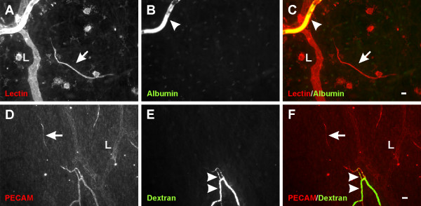
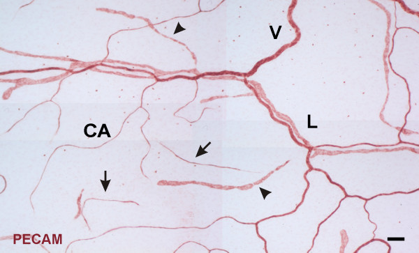
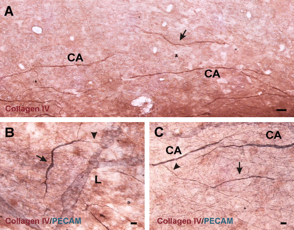
Results: Mesenteric tissues, which allow visualization of entire microvascular networks at a single cell level, were harvested from unstimulated adult male Wistar rats and Wistar rats 3 and 10 days post angiogenesis stimulation by mast cell degranulation with compound 48/80. Tissues were immunolabeled for PECAM and BRDU. Identification of vessel lumens via injection of FITC-dextran confirmed that endothelial cell segments were disconnected from nearby patent networks. Stimulated networks displayed increases in vascular area, length density, and capillary sprouting. On day 3, the percentage of islands with at least one BRDU-positive cell increased compared to the unstimulated level and was equal to the percentage of capillary sprouts with at least one BRDU-positive cell. At day 10, the number of vascular islands per vascular area dramatically decreased compared to unstimulated and day 3 levels.
Conclusions: These results show that vascular islands have the ability to proliferate and suggest that they are able to incorporate into the microcirculation during the initial stages of microvascular network growth.
BMC PhysiologyBiochemistry, Genetics and Molecular Biology-Physiology
CiteScore
9.60
自引率
0.00%
发文量
0
期刊介绍:
BMC Physiology is an open access journal publishing original peer-reviewed research articles in cellular, tissue-level, organismal, functional, and developmental aspects of physiological processes. BMC Physiology (ISSN 1472-6793) is indexed/tracked/covered by PubMed, MEDLINE, BIOSIS, CAS, EMBASE, Scopus, Zoological Record and Google Scholar.


 分享
分享
 求助内容:
求助内容: 应助结果提醒方式:
 扫码关注我们
