Subhodeep Moitra, Kalyan C Tirupula, Judith Klein-Seetharaman, Christopher James Langmead
{"title":"通过对G蛋白偶联受体的多序列和结构分析,确定了相关突变网络中的最小配体结合袋。","authors":"Subhodeep Moitra, Kalyan C Tirupula, Judith Klein-Seetharaman, Christopher James Langmead","doi":"10.1186/2046-1682-5-13","DOIUrl":null,"url":null,"abstract":"<p><strong>Background: </strong>G protein coupled receptors (GPCRs) are seven helical transmembrane proteins that function as signal transducers. They bind ligands in their extracellular and transmembrane regions and activate cognate G proteins at their intracellular surface at the other side of the membrane. The relay of allosteric communication between the ligand binding site and the distant G protein binding site is poorly understood. In this study, GREMLIN 1, a recently developed method that identifies networks of co-evolving residues from multiple sequence alignments, was used to identify those that may be involved in communicating the activation signal across the membrane. The GREMLIN-predicted long-range interactions between amino acids were analyzed with respect to the seven GPCR structures that have been crystallized at the time this study was undertaken.</p><p><strong>Results: </strong>GREMLIN significantly enriches the edges containing residues that are part of the ligand binding pocket, when compared to a control distribution of edges drawn from a random graph. An analysis of these edges reveals a minimal GPCR binding pocket containing four residues (T1183.33, M2075.42, Y2686.51 and A2927.39). Additionally, of the ten residues predicted to have the most long-range interactions (A1173.32, A2726.55, E1133.28, H2115.46, S186EC2, A2927.39, E1223.37, G902.57, G1143.29 and M2075.42), nine are part of the ligand binding pocket.</p><p><strong>Conclusions: </strong>We demonstrate the use of GREMLIN to reveal a network of statistically correlated and functionally important residues in class A GPCRs. GREMLIN identified that ligand binding pocket residues are extensively correlated with distal residues. An analysis of the GREMLIN edges across multiple structures suggests that there may be a minimal binding pocket common to the seven known GPCRs. Further, the activation of rhodopsin involves these long-range interactions between extracellular and intracellular domain residues mediated by the retinal domain.</p>","PeriodicalId":9045,"journal":{"name":"BMC Biophysics","volume":"5 ","pages":"13"},"PeriodicalIF":0.0000,"publicationDate":"2012-06-29","publicationTypes":"Journal Article","fieldsOfStudy":null,"isOpenAccess":false,"openAccessPdf":"https://sci-hub-pdf.com/10.1186/2046-1682-5-13","citationCount":"8","resultStr":"{\"title\":\"A minimal ligand binding pocket within a network of correlated mutations identified by multiple sequence and structural analysis of G protein coupled receptors.\",\"authors\":\"Subhodeep Moitra, Kalyan C Tirupula, Judith Klein-Seetharaman, Christopher James Langmead\",\"doi\":\"10.1186/2046-1682-5-13\",\"DOIUrl\":null,\"url\":null,\"abstract\":\"<p><strong>Background: </strong>G protein coupled receptors (GPCRs) are seven helical transmembrane proteins that function as signal transducers. They bind ligands in their extracellular and transmembrane regions and activate cognate G proteins at their intracellular surface at the other side of the membrane. The relay of allosteric communication between the ligand binding site and the distant G protein binding site is poorly understood. In this study, GREMLIN 1, a recently developed method that identifies networks of co-evolving residues from multiple sequence alignments, was used to identify those that may be involved in communicating the activation signal across the membrane. The GREMLIN-predicted long-range interactions between amino acids were analyzed with respect to the seven GPCR structures that have been crystallized at the time this study was undertaken.</p><p><strong>Results: </strong>GREMLIN significantly enriches the edges containing residues that are part of the ligand binding pocket, when compared to a control distribution of edges drawn from a random graph. An analysis of these edges reveals a minimal GPCR binding pocket containing four residues (T1183.33, M2075.42, Y2686.51 and A2927.39). Additionally, of the ten residues predicted to have the most long-range interactions (A1173.32, A2726.55, E1133.28, H2115.46, S186EC2, A2927.39, E1223.37, G902.57, G1143.29 and M2075.42), nine are part of the ligand binding pocket.</p><p><strong>Conclusions: </strong>We demonstrate the use of GREMLIN to reveal a network of statistically correlated and functionally important residues in class A GPCRs. GREMLIN identified that ligand binding pocket residues are extensively correlated with distal residues. An analysis of the GREMLIN edges across multiple structures suggests that there may be a minimal binding pocket common to the seven known GPCRs. Further, the activation of rhodopsin involves these long-range interactions between extracellular and intracellular domain residues mediated by the retinal domain.</p>\",\"PeriodicalId\":9045,\"journal\":{\"name\":\"BMC Biophysics\",\"volume\":\"5 \",\"pages\":\"13\"},\"PeriodicalIF\":0.0000,\"publicationDate\":\"2012-06-29\",\"publicationTypes\":\"Journal Article\",\"fieldsOfStudy\":null,\"isOpenAccess\":false,\"openAccessPdf\":\"https://sci-hub-pdf.com/10.1186/2046-1682-5-13\",\"citationCount\":\"8\",\"resultStr\":null,\"platform\":\"Semanticscholar\",\"paperid\":null,\"PeriodicalName\":\"BMC Biophysics\",\"FirstCategoryId\":\"1085\",\"ListUrlMain\":\"https://doi.org/10.1186/2046-1682-5-13\",\"RegionNum\":0,\"RegionCategory\":null,\"ArticlePicture\":[],\"TitleCN\":null,\"AbstractTextCN\":null,\"PMCID\":null,\"EPubDate\":\"\",\"PubModel\":\"\",\"JCR\":\"Q1\",\"JCRName\":\"Biochemistry, Genetics and Molecular Biology\",\"Score\":null,\"Total\":0}","platform":"Semanticscholar","paperid":null,"PeriodicalName":"BMC Biophysics","FirstCategoryId":"1085","ListUrlMain":"https://doi.org/10.1186/2046-1682-5-13","RegionNum":0,"RegionCategory":null,"ArticlePicture":[],"TitleCN":null,"AbstractTextCN":null,"PMCID":null,"EPubDate":"","PubModel":"","JCR":"Q1","JCRName":"Biochemistry, Genetics and Molecular Biology","Score":null,"Total":0}
A minimal ligand binding pocket within a network of correlated mutations identified by multiple sequence and structural analysis of G protein coupled receptors.
Background: G protein coupled receptors (GPCRs) are seven helical transmembrane proteins that function as signal transducers. They bind ligands in their extracellular and transmembrane regions and activate cognate G proteins at their intracellular surface at the other side of the membrane. The relay of allosteric communication between the ligand binding site and the distant G protein binding site is poorly understood. In this study, GREMLIN 1, a recently developed method that identifies networks of co-evolving residues from multiple sequence alignments, was used to identify those that may be involved in communicating the activation signal across the membrane. The GREMLIN-predicted long-range interactions between amino acids were analyzed with respect to the seven GPCR structures that have been crystallized at the time this study was undertaken.
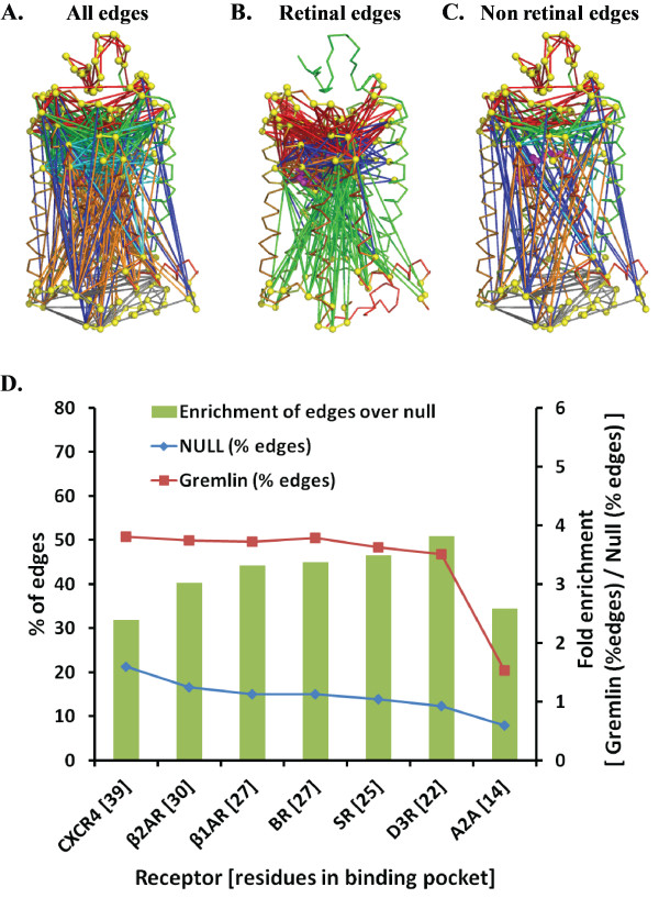
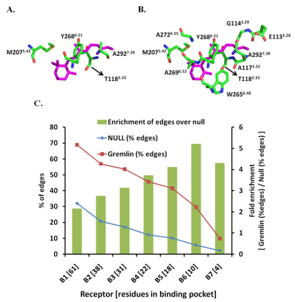
Results: GREMLIN significantly enriches the edges containing residues that are part of the ligand binding pocket, when compared to a control distribution of edges drawn from a random graph. An analysis of these edges reveals a minimal GPCR binding pocket containing four residues (T1183.33, M2075.42, Y2686.51 and A2927.39). Additionally, of the ten residues predicted to have the most long-range interactions (A1173.32, A2726.55, E1133.28, H2115.46, S186EC2, A2927.39, E1223.37, G902.57, G1143.29 and M2075.42), nine are part of the ligand binding pocket.
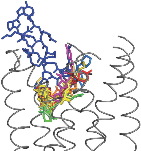
Conclusions: We demonstrate the use of GREMLIN to reveal a network of statistically correlated and functionally important residues in class A GPCRs. GREMLIN identified that ligand binding pocket residues are extensively correlated with distal residues. An analysis of the GREMLIN edges across multiple structures suggests that there may be a minimal binding pocket common to the seven known GPCRs. Further, the activation of rhodopsin involves these long-range interactions between extracellular and intracellular domain residues mediated by the retinal domain.


 分享
分享
 求助内容:
求助内容: 应助结果提醒方式:
 扫码关注我们
