Chandra Kirana, Hongjun Shi, Emma Laing, Kylie Hood, Rose Miller, Peter Bethwaite, John Keating, T William Jordan, Mark Hayes, Richard Stubbs
{"title":"组织蛋白酶D在结直肠癌中的表达:通过Western Blotting、免疫组织化学和组织微阵列验证的蛋白质组学发现。","authors":"Chandra Kirana, Hongjun Shi, Emma Laing, Kylie Hood, Rose Miller, Peter Bethwaite, John Keating, T William Jordan, Mark Hayes, Richard Stubbs","doi":"10.1155/2012/245819","DOIUrl":null,"url":null,"abstract":"<p><p>Despite recent advances in surgical techniques and therapeutic treatments, survival from colorectal cancer (CRC) remains disappointing with some 40-50% of newly diagnosed patients ultimately dying of metastatic disease. Current staging by light microscopy alone is not sufficiently predictive of prognosis and would benefit from additional support from biomarkers in order to stratify patients appropriately for adjuvant therapy. We have identified that cathepsin D expression was significantly greater in cells from invasive front (IF) area and liver metastasis (LM) than those from main tumour body (MTB). Cathepsin D expression was subsequently examined by immunohistochemistry in tissue microarrays from 119 patients with CRC. Strong expression in tumour cells at the IF did not correlate significantly with any clinico-pathological parameters examined or patient survival. However, cathepsin D expression in cells from the MTB was highly elevated in late stage CRC and showed significant correlation with subsequent distant metastasis and shorter cancer-specific survival. We also found that macrophages surrounding tumour cells stained strongly for cathepsin D but there was no significant correlation found between cathepsin D in macrophages at IF and MTB of CRC patient with the clinic-pathological parameters examined.</p>","PeriodicalId":73474,"journal":{"name":"International journal of proteomics","volume":"2012 ","pages":"245819"},"PeriodicalIF":0.0000,"publicationDate":"2012-01-01","publicationTypes":"Journal Article","fieldsOfStudy":null,"isOpenAccess":false,"openAccessPdf":"https://sci-hub-pdf.com/10.1155/2012/245819","citationCount":"32","resultStr":"{\"title\":\"Cathepsin D Expression in Colorectal Cancer: From Proteomic Discovery through Validation Using Western Blotting, Immunohistochemistry, and Tissue Microarrays.\",\"authors\":\"Chandra Kirana, Hongjun Shi, Emma Laing, Kylie Hood, Rose Miller, Peter Bethwaite, John Keating, T William Jordan, Mark Hayes, Richard Stubbs\",\"doi\":\"10.1155/2012/245819\",\"DOIUrl\":null,\"url\":null,\"abstract\":\"<p><p>Despite recent advances in surgical techniques and therapeutic treatments, survival from colorectal cancer (CRC) remains disappointing with some 40-50% of newly diagnosed patients ultimately dying of metastatic disease. Current staging by light microscopy alone is not sufficiently predictive of prognosis and would benefit from additional support from biomarkers in order to stratify patients appropriately for adjuvant therapy. We have identified that cathepsin D expression was significantly greater in cells from invasive front (IF) area and liver metastasis (LM) than those from main tumour body (MTB). Cathepsin D expression was subsequently examined by immunohistochemistry in tissue microarrays from 119 patients with CRC. Strong expression in tumour cells at the IF did not correlate significantly with any clinico-pathological parameters examined or patient survival. However, cathepsin D expression in cells from the MTB was highly elevated in late stage CRC and showed significant correlation with subsequent distant metastasis and shorter cancer-specific survival. We also found that macrophages surrounding tumour cells stained strongly for cathepsin D but there was no significant correlation found between cathepsin D in macrophages at IF and MTB of CRC patient with the clinic-pathological parameters examined.</p>\",\"PeriodicalId\":73474,\"journal\":{\"name\":\"International journal of proteomics\",\"volume\":\"2012 \",\"pages\":\"245819\"},\"PeriodicalIF\":0.0000,\"publicationDate\":\"2012-01-01\",\"publicationTypes\":\"Journal Article\",\"fieldsOfStudy\":null,\"isOpenAccess\":false,\"openAccessPdf\":\"https://sci-hub-pdf.com/10.1155/2012/245819\",\"citationCount\":\"32\",\"resultStr\":null,\"platform\":\"Semanticscholar\",\"paperid\":null,\"PeriodicalName\":\"International journal of proteomics\",\"FirstCategoryId\":\"1085\",\"ListUrlMain\":\"https://doi.org/10.1155/2012/245819\",\"RegionNum\":0,\"RegionCategory\":null,\"ArticlePicture\":[],\"TitleCN\":null,\"AbstractTextCN\":null,\"PMCID\":null,\"EPubDate\":\"2012/8/7 0:00:00\",\"PubModel\":\"Epub\",\"JCR\":\"\",\"JCRName\":\"\",\"Score\":null,\"Total\":0}","platform":"Semanticscholar","paperid":null,"PeriodicalName":"International journal of proteomics","FirstCategoryId":"1085","ListUrlMain":"https://doi.org/10.1155/2012/245819","RegionNum":0,"RegionCategory":null,"ArticlePicture":[],"TitleCN":null,"AbstractTextCN":null,"PMCID":null,"EPubDate":"2012/8/7 0:00:00","PubModel":"Epub","JCR":"","JCRName":"","Score":null,"Total":0}
Cathepsin D Expression in Colorectal Cancer: From Proteomic Discovery through Validation Using Western Blotting, Immunohistochemistry, and Tissue Microarrays.
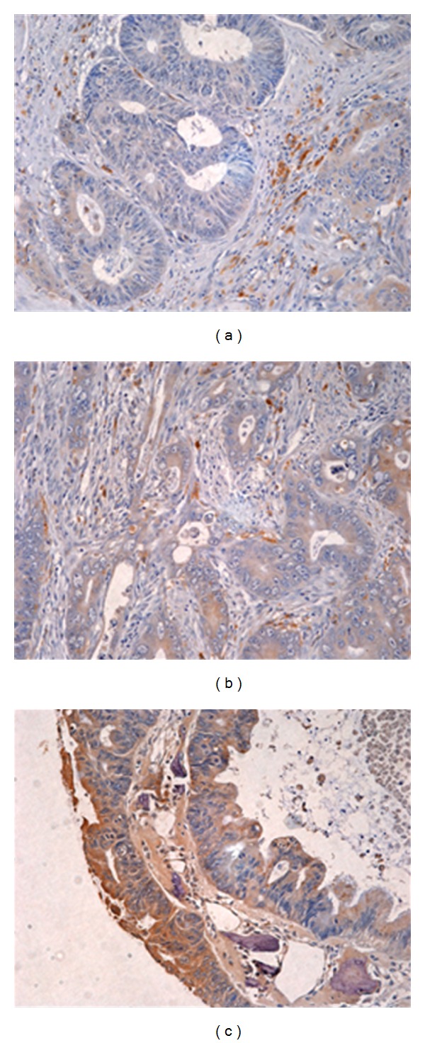
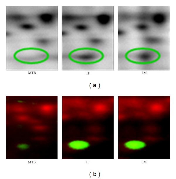
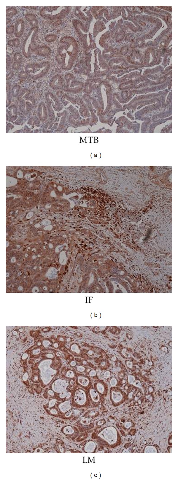
Despite recent advances in surgical techniques and therapeutic treatments, survival from colorectal cancer (CRC) remains disappointing with some 40-50% of newly diagnosed patients ultimately dying of metastatic disease. Current staging by light microscopy alone is not sufficiently predictive of prognosis and would benefit from additional support from biomarkers in order to stratify patients appropriately for adjuvant therapy. We have identified that cathepsin D expression was significantly greater in cells from invasive front (IF) area and liver metastasis (LM) than those from main tumour body (MTB). Cathepsin D expression was subsequently examined by immunohistochemistry in tissue microarrays from 119 patients with CRC. Strong expression in tumour cells at the IF did not correlate significantly with any clinico-pathological parameters examined or patient survival. However, cathepsin D expression in cells from the MTB was highly elevated in late stage CRC and showed significant correlation with subsequent distant metastasis and shorter cancer-specific survival. We also found that macrophages surrounding tumour cells stained strongly for cathepsin D but there was no significant correlation found between cathepsin D in macrophages at IF and MTB of CRC patient with the clinic-pathological parameters examined.


 分享
分享

 求助内容:
求助内容: 应助结果提醒方式:
 扫码关注我们
