Andreas S Schneider, Birgit Heiland, Nicolas J Peter, Christina Guth, Eduard Arzt, Ingrid M Weiss
{"title":"偏振光显微镜、电子显微镜和纳米压痕鉴定的分层超结构:生物控制对鲍鱼海壳生长模式的限制意义。","authors":"Andreas S Schneider, Birgit Heiland, Nicolas J Peter, Christina Guth, Eduard Arzt, Ingrid M Weiss","doi":"10.1186/2046-1682-5-19","DOIUrl":null,"url":null,"abstract":"<p><strong>Unlabelled: </strong></p><p><strong>Background: </strong>Mollusc shells are commonly investigated using high-resolution imaging techniques based on cryo-fixation. Less detailed information is available regarding the light-optical properties. Sea shells of Haliotis pulcherina were embedded for polishing in defined orientations in order to investigate the interface between prismatic calcite and nacreous aragonite by standard materialographic methods. A polished thin section of the interface was prepared with a defined thickness of 60 μm for quantitative birefringence analysis using polarized light and LC-PolScope microscopy. Scanning electron microscopy images were obtained for comparison. In order to study structural-mechanical relationships, nanoindentation experiments were performed.</p><p><strong>Results: </strong>Incident light microscopy revealed a super-structure in semi-transparent regions of the polished cross-section under a defined angle. This super-structure is not visible in transmitted birefringence analysis due to the blurred polarization of small nacre platelets and numerous organic interfaces. The relative orientation and homogeneity of calcite prisms was directly identified, some of them with their optical axes exactly normal to the imaging plane. Co-oriented \"prism colonies\" were identified by polarized light analyses. The nacreous super-structure was also visualized by secondary electron imaging under defined angles. The domains of the super-structure were interpreted to consist of crystallographically aligned platelet stacks. Nanoindentation experiments showed that mechanical properties changed with the same periodicity as the domain size.</p><p><strong>Conclusions: </strong>In this study, we have demonstrated that insights into the growth mechanisms of nacre can be obtained by conventional light-optical methods. For example, we observed super-structures formed by co-oriented nacre platelets as previously identified using X-ray Photo-electron Emission Microscopy (X-PEEM) [Gilbert et al., Journal of the American Chemical Society 2008, 130:17519-17527]. Polarized optical microscopy revealed unprecedented super-structures in the calcitic shell part. This bears, in principle, the potential for in vivo studies, which might be useful for investigating the growth modes of nacre and other shell types.</p>","PeriodicalId":9045,"journal":{"name":"BMC Biophysics","volume":"5 ","pages":"19"},"PeriodicalIF":0.0000,"publicationDate":"2012-09-12","publicationTypes":"Journal Article","fieldsOfStudy":null,"isOpenAccess":false,"openAccessPdf":"https://sci-hub-pdf.com/10.1186/2046-1682-5-19","citationCount":"19","resultStr":"{\"title\":\"Hierarchical super-structure identified by polarized light microscopy, electron microscopy and nanoindentation: Implications for the limits of biological control over the growth mode of abalone sea shells.\",\"authors\":\"Andreas S Schneider, Birgit Heiland, Nicolas J Peter, Christina Guth, Eduard Arzt, Ingrid M Weiss\",\"doi\":\"10.1186/2046-1682-5-19\",\"DOIUrl\":null,\"url\":null,\"abstract\":\"<p><strong>Unlabelled: </strong></p><p><strong>Background: </strong>Mollusc shells are commonly investigated using high-resolution imaging techniques based on cryo-fixation. Less detailed information is available regarding the light-optical properties. Sea shells of Haliotis pulcherina were embedded for polishing in defined orientations in order to investigate the interface between prismatic calcite and nacreous aragonite by standard materialographic methods. A polished thin section of the interface was prepared with a defined thickness of 60 μm for quantitative birefringence analysis using polarized light and LC-PolScope microscopy. Scanning electron microscopy images were obtained for comparison. In order to study structural-mechanical relationships, nanoindentation experiments were performed.</p><p><strong>Results: </strong>Incident light microscopy revealed a super-structure in semi-transparent regions of the polished cross-section under a defined angle. This super-structure is not visible in transmitted birefringence analysis due to the blurred polarization of small nacre platelets and numerous organic interfaces. The relative orientation and homogeneity of calcite prisms was directly identified, some of them with their optical axes exactly normal to the imaging plane. Co-oriented \\\"prism colonies\\\" were identified by polarized light analyses. The nacreous super-structure was also visualized by secondary electron imaging under defined angles. The domains of the super-structure were interpreted to consist of crystallographically aligned platelet stacks. Nanoindentation experiments showed that mechanical properties changed with the same periodicity as the domain size.</p><p><strong>Conclusions: </strong>In this study, we have demonstrated that insights into the growth mechanisms of nacre can be obtained by conventional light-optical methods. For example, we observed super-structures formed by co-oriented nacre platelets as previously identified using X-ray Photo-electron Emission Microscopy (X-PEEM) [Gilbert et al., Journal of the American Chemical Society 2008, 130:17519-17527]. Polarized optical microscopy revealed unprecedented super-structures in the calcitic shell part. This bears, in principle, the potential for in vivo studies, which might be useful for investigating the growth modes of nacre and other shell types.</p>\",\"PeriodicalId\":9045,\"journal\":{\"name\":\"BMC Biophysics\",\"volume\":\"5 \",\"pages\":\"19\"},\"PeriodicalIF\":0.0000,\"publicationDate\":\"2012-09-12\",\"publicationTypes\":\"Journal Article\",\"fieldsOfStudy\":null,\"isOpenAccess\":false,\"openAccessPdf\":\"https://sci-hub-pdf.com/10.1186/2046-1682-5-19\",\"citationCount\":\"19\",\"resultStr\":null,\"platform\":\"Semanticscholar\",\"paperid\":null,\"PeriodicalName\":\"BMC Biophysics\",\"FirstCategoryId\":\"1085\",\"ListUrlMain\":\"https://doi.org/10.1186/2046-1682-5-19\",\"RegionNum\":0,\"RegionCategory\":null,\"ArticlePicture\":[],\"TitleCN\":null,\"AbstractTextCN\":null,\"PMCID\":null,\"EPubDate\":\"\",\"PubModel\":\"\",\"JCR\":\"Q1\",\"JCRName\":\"Biochemistry, Genetics and Molecular Biology\",\"Score\":null,\"Total\":0}","platform":"Semanticscholar","paperid":null,"PeriodicalName":"BMC Biophysics","FirstCategoryId":"1085","ListUrlMain":"https://doi.org/10.1186/2046-1682-5-19","RegionNum":0,"RegionCategory":null,"ArticlePicture":[],"TitleCN":null,"AbstractTextCN":null,"PMCID":null,"EPubDate":"","PubModel":"","JCR":"Q1","JCRName":"Biochemistry, Genetics and Molecular Biology","Score":null,"Total":0}
引用次数: 19
摘要
背景:软体动物壳通常使用基于冷冻固定的高分辨率成像技术进行研究。关于光光学性质的详细信息较少。为了用标准的材料学方法研究柱状方解石与珠光文石之间的界面,对pulcherina海螺壳进行了定向镶嵌抛光。制备了厚度为60 μm的抛光界面薄片,利用偏振光和LC-PolScope显微镜进行定量双折射分析。扫描电镜图像进行比较。为了研究结构-力学关系,进行了纳米压痕实验。结果:入射光显微镜显示,在一定角度下,抛光截面的半透明区域有超结构。这种超结构在透射双折射分析中是不可见的,因为小珠层和许多有机界面的偏振模糊。直接识别了方解石棱镜的相对取向和均匀性,其中一些方解石棱镜的光轴与成像平面完全垂直。通过偏振光分析确定了共取向的“棱镜菌落”。在确定的角度下,通过二次电子成像显示了珍珠层的上部结构。超结构的畴被解释为由晶体学上排列的血小板堆叠组成。纳米压痕实验表明,材料的力学性能随畴尺寸的变化具有相同的周期性。结论:在本研究中,我们证明了通过传统的光光学方法可以深入了解珍珠层的生长机制。例如,我们通过x射线光电子发射显微镜(X-PEEM)观察到由共取向珍珠层血小板形成的超结构[Gilbert等人,Journal of American Chemical Society 2008, 130:17519-17527]。偏光显微镜显示,在钙质外壳部分前所未有的超结构。原则上,这有可能在体内进行研究,这可能对研究珍珠层和其他贝壳类型的生长模式有用。
Hierarchical super-structure identified by polarized light microscopy, electron microscopy and nanoindentation: Implications for the limits of biological control over the growth mode of abalone sea shells.
Unlabelled:
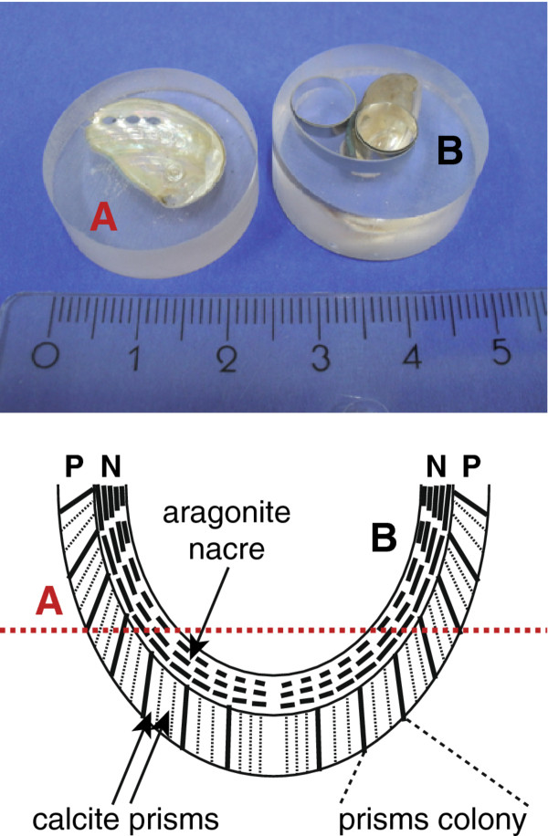
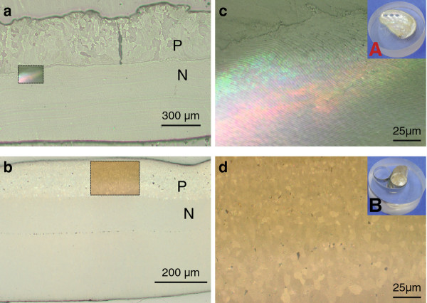
Background: Mollusc shells are commonly investigated using high-resolution imaging techniques based on cryo-fixation. Less detailed information is available regarding the light-optical properties. Sea shells of Haliotis pulcherina were embedded for polishing in defined orientations in order to investigate the interface between prismatic calcite and nacreous aragonite by standard materialographic methods. A polished thin section of the interface was prepared with a defined thickness of 60 μm for quantitative birefringence analysis using polarized light and LC-PolScope microscopy. Scanning electron microscopy images were obtained for comparison. In order to study structural-mechanical relationships, nanoindentation experiments were performed.
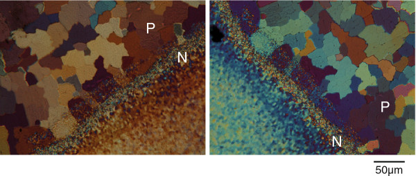
Results: Incident light microscopy revealed a super-structure in semi-transparent regions of the polished cross-section under a defined angle. This super-structure is not visible in transmitted birefringence analysis due to the blurred polarization of small nacre platelets and numerous organic interfaces. The relative orientation and homogeneity of calcite prisms was directly identified, some of them with their optical axes exactly normal to the imaging plane. Co-oriented "prism colonies" were identified by polarized light analyses. The nacreous super-structure was also visualized by secondary electron imaging under defined angles. The domains of the super-structure were interpreted to consist of crystallographically aligned platelet stacks. Nanoindentation experiments showed that mechanical properties changed with the same periodicity as the domain size.
Conclusions: In this study, we have demonstrated that insights into the growth mechanisms of nacre can be obtained by conventional light-optical methods. For example, we observed super-structures formed by co-oriented nacre platelets as previously identified using X-ray Photo-electron Emission Microscopy (X-PEEM) [Gilbert et al., Journal of the American Chemical Society 2008, 130:17519-17527]. Polarized optical microscopy revealed unprecedented super-structures in the calcitic shell part. This bears, in principle, the potential for in vivo studies, which might be useful for investigating the growth modes of nacre and other shell types.


 分享
分享
 求助内容:
求助内容: 应助结果提醒方式:
 扫码关注我们
