{"title":"TRPV4通道在人收集管细胞中的功能表达:对糖尿病肾病继发性高血压的影响","authors":"Claire E Hills, Rosemary Bland, Paul E Squires","doi":"10.1155/2012/936518","DOIUrl":null,"url":null,"abstract":"<p><strong>Background: </strong>The Vanilloid subfamily of transient receptor potential (TRPV) ion channels has been widely implicated in detecting osmotic and mechanical stress. In the current study, we examine the functional expression of TRPV4 channels in cell volume regulation in cells of the human collecting duct.</p><p><strong>Methods: </strong>Western blot analysis, siRNA knockdown, and microfluorimetry were used to assess the expression and function of TRPV4 in mediating Ca²⁺-dependent mechanical stimulation within a novel system of the human collecting duct (HCD).</p><p><strong>Results: </strong>Native and siRNA knockdown of TRPV4 protein expression was confirmed by western blot analysis. Touch was used as a cell-directed surrogate for osmotic stress. Mechanical stimulation of HCD cells evoked a transient increase in [Ca²⁺](i) that was dependent upon thapsigargin-sensitive store release and Ca²⁺ influx. At 48 hrs, high glucose and mannitol (25 mM) reduced TRPV4 expression by 54% and 24%, respectively. Similar treatment doubled SGK1 expression. Touch-evoked changes were negated following TRPV4 knockdown.</p><p><strong>Conclusion: </strong>Our data confirm expression of Ca²⁺-dependent TRPV4 channels in HCD cells and suggest that a loss of expression in response to high glucose attenuates the ability of the collecting duct to exhibit regulatory volume decreases, an effect that may contribute to the pathology of fluid and electrolyte imbalance as observed in diabetic nephropathy.</p>","PeriodicalId":12109,"journal":{"name":"Experimental Diabetes Research","volume":"2012 ","pages":"936518"},"PeriodicalIF":0.0000,"publicationDate":"2012-01-01","publicationTypes":"Journal Article","fieldsOfStudy":null,"isOpenAccess":false,"openAccessPdf":"https://sci-hub-pdf.com/10.1155/2012/936518","citationCount":"21","resultStr":"{\"title\":\"Functional expression of TRPV4 channels in human collecting duct cells: implications for secondary hypertension in diabetic nephropathy.\",\"authors\":\"Claire E Hills, Rosemary Bland, Paul E Squires\",\"doi\":\"10.1155/2012/936518\",\"DOIUrl\":null,\"url\":null,\"abstract\":\"<p><strong>Background: </strong>The Vanilloid subfamily of transient receptor potential (TRPV) ion channels has been widely implicated in detecting osmotic and mechanical stress. In the current study, we examine the functional expression of TRPV4 channels in cell volume regulation in cells of the human collecting duct.</p><p><strong>Methods: </strong>Western blot analysis, siRNA knockdown, and microfluorimetry were used to assess the expression and function of TRPV4 in mediating Ca²⁺-dependent mechanical stimulation within a novel system of the human collecting duct (HCD).</p><p><strong>Results: </strong>Native and siRNA knockdown of TRPV4 protein expression was confirmed by western blot analysis. Touch was used as a cell-directed surrogate for osmotic stress. Mechanical stimulation of HCD cells evoked a transient increase in [Ca²⁺](i) that was dependent upon thapsigargin-sensitive store release and Ca²⁺ influx. At 48 hrs, high glucose and mannitol (25 mM) reduced TRPV4 expression by 54% and 24%, respectively. Similar treatment doubled SGK1 expression. Touch-evoked changes were negated following TRPV4 knockdown.</p><p><strong>Conclusion: </strong>Our data confirm expression of Ca²⁺-dependent TRPV4 channels in HCD cells and suggest that a loss of expression in response to high glucose attenuates the ability of the collecting duct to exhibit regulatory volume decreases, an effect that may contribute to the pathology of fluid and electrolyte imbalance as observed in diabetic nephropathy.</p>\",\"PeriodicalId\":12109,\"journal\":{\"name\":\"Experimental Diabetes Research\",\"volume\":\"2012 \",\"pages\":\"936518\"},\"PeriodicalIF\":0.0000,\"publicationDate\":\"2012-01-01\",\"publicationTypes\":\"Journal Article\",\"fieldsOfStudy\":null,\"isOpenAccess\":false,\"openAccessPdf\":\"https://sci-hub-pdf.com/10.1155/2012/936518\",\"citationCount\":\"21\",\"resultStr\":null,\"platform\":\"Semanticscholar\",\"paperid\":null,\"PeriodicalName\":\"Experimental Diabetes Research\",\"FirstCategoryId\":\"1085\",\"ListUrlMain\":\"https://doi.org/10.1155/2012/936518\",\"RegionNum\":0,\"RegionCategory\":null,\"ArticlePicture\":[],\"TitleCN\":null,\"AbstractTextCN\":null,\"PMCID\":null,\"EPubDate\":\"2012/9/20 0:00:00\",\"PubModel\":\"Epub\",\"JCR\":\"\",\"JCRName\":\"\",\"Score\":null,\"Total\":0}","platform":"Semanticscholar","paperid":null,"PeriodicalName":"Experimental Diabetes Research","FirstCategoryId":"1085","ListUrlMain":"https://doi.org/10.1155/2012/936518","RegionNum":0,"RegionCategory":null,"ArticlePicture":[],"TitleCN":null,"AbstractTextCN":null,"PMCID":null,"EPubDate":"2012/9/20 0:00:00","PubModel":"Epub","JCR":"","JCRName":"","Score":null,"Total":0}
Functional expression of TRPV4 channels in human collecting duct cells: implications for secondary hypertension in diabetic nephropathy.
Background: The Vanilloid subfamily of transient receptor potential (TRPV) ion channels has been widely implicated in detecting osmotic and mechanical stress. In the current study, we examine the functional expression of TRPV4 channels in cell volume regulation in cells of the human collecting duct.
Methods: Western blot analysis, siRNA knockdown, and microfluorimetry were used to assess the expression and function of TRPV4 in mediating Ca²⁺-dependent mechanical stimulation within a novel system of the human collecting duct (HCD).
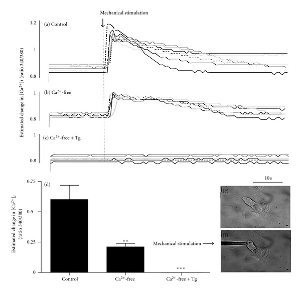
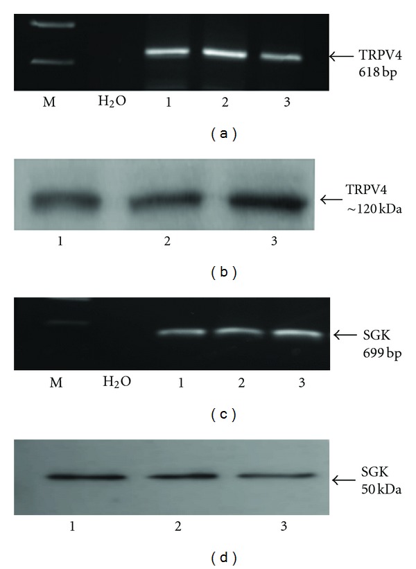
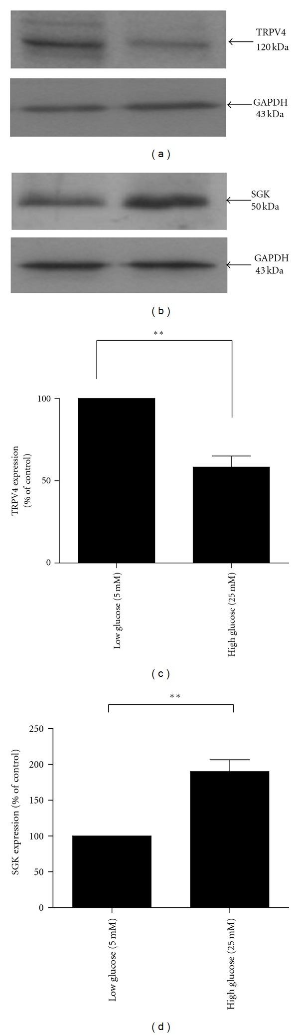
Results: Native and siRNA knockdown of TRPV4 protein expression was confirmed by western blot analysis. Touch was used as a cell-directed surrogate for osmotic stress. Mechanical stimulation of HCD cells evoked a transient increase in [Ca²⁺](i) that was dependent upon thapsigargin-sensitive store release and Ca²⁺ influx. At 48 hrs, high glucose and mannitol (25 mM) reduced TRPV4 expression by 54% and 24%, respectively. Similar treatment doubled SGK1 expression. Touch-evoked changes were negated following TRPV4 knockdown.
Conclusion: Our data confirm expression of Ca²⁺-dependent TRPV4 channels in HCD cells and suggest that a loss of expression in response to high glucose attenuates the ability of the collecting duct to exhibit regulatory volume decreases, an effect that may contribute to the pathology of fluid and electrolyte imbalance as observed in diabetic nephropathy.


 分享
分享
 求助内容:
求助内容: 应助结果提醒方式:
 扫码关注我们
