{"title":"马羊膜间充质干细胞的分离与鉴定","authors":"Alessandra Coli, Francesca Nocchi, Roberta Lamanna, Mariacarla Iorio, Simone Lapi, Patrizia Urciuoli, Fabrizio Scatena, Elisabetta Giannessi, Maria Rita Stornelli, Simona Passeri","doi":"10.1042/CBR20110004","DOIUrl":null,"url":null,"abstract":"<p>The amnion is a particular tissue whose cells show features of multipotent stem cells proposed for use in cellular therapy and regenerative medicine. From equine amnion collected after the foal birth we have isolated MSCs (mesenchymal stem cells), namely EAMSCs (equine amnion mesenchymal stem cells), from the mesoblastic layer. The cells were grown in α-MEM (α-modified minimum essential medium) and the effect of EGF (epidermal growth factor) supplementation was evaluated. To assess the growth kinetic of EAMSCs we have taken into account some parameters [PD (population doubling), fold increase and DT (doubling time)]. The differentiation in chondrogenic, adipogenic and osteogenic types of cells and their epitope expression by a cytofluorimetric study have been reported. EGF supplementation of the culture medium resulted in a significant increase in PD growth parameter and in the formation of bone nodules for the osteogenic differentiation. By immunohistochemistry the amnion tissue shows a positivity for the c-Kit (cluster tyrosine-protein kinase), CD105 and Oct-4 (octamer-binding transcription factor 4) antigens that confirmed the presence of MSCs with embryonic phenotype.</p>","PeriodicalId":75683,"journal":{"name":"Cell biology international reports","volume":"18 1","pages":"23-29"},"PeriodicalIF":0.0000,"publicationDate":"2013-06-25","publicationTypes":"Journal Article","fieldsOfStudy":null,"isOpenAccess":false,"openAccessPdf":"https://sci-hub-pdf.com/10.1042/CBR20110004","citationCount":"22","resultStr":"{\"title\":\"Isolation and characterization of equine amnion mesenchymal stem cells\",\"authors\":\"Alessandra Coli, Francesca Nocchi, Roberta Lamanna, Mariacarla Iorio, Simone Lapi, Patrizia Urciuoli, Fabrizio Scatena, Elisabetta Giannessi, Maria Rita Stornelli, Simona Passeri\",\"doi\":\"10.1042/CBR20110004\",\"DOIUrl\":null,\"url\":null,\"abstract\":\"<p>The amnion is a particular tissue whose cells show features of multipotent stem cells proposed for use in cellular therapy and regenerative medicine. From equine amnion collected after the foal birth we have isolated MSCs (mesenchymal stem cells), namely EAMSCs (equine amnion mesenchymal stem cells), from the mesoblastic layer. The cells were grown in α-MEM (α-modified minimum essential medium) and the effect of EGF (epidermal growth factor) supplementation was evaluated. To assess the growth kinetic of EAMSCs we have taken into account some parameters [PD (population doubling), fold increase and DT (doubling time)]. The differentiation in chondrogenic, adipogenic and osteogenic types of cells and their epitope expression by a cytofluorimetric study have been reported. EGF supplementation of the culture medium resulted in a significant increase in PD growth parameter and in the formation of bone nodules for the osteogenic differentiation. By immunohistochemistry the amnion tissue shows a positivity for the c-Kit (cluster tyrosine-protein kinase), CD105 and Oct-4 (octamer-binding transcription factor 4) antigens that confirmed the presence of MSCs with embryonic phenotype.</p>\",\"PeriodicalId\":75683,\"journal\":{\"name\":\"Cell biology international reports\",\"volume\":\"18 1\",\"pages\":\"23-29\"},\"PeriodicalIF\":0.0000,\"publicationDate\":\"2013-06-25\",\"publicationTypes\":\"Journal Article\",\"fieldsOfStudy\":null,\"isOpenAccess\":false,\"openAccessPdf\":\"https://sci-hub-pdf.com/10.1042/CBR20110004\",\"citationCount\":\"22\",\"resultStr\":null,\"platform\":\"Semanticscholar\",\"paperid\":null,\"PeriodicalName\":\"Cell biology international reports\",\"FirstCategoryId\":\"1085\",\"ListUrlMain\":\"https://onlinelibrary.wiley.com/doi/10.1042/CBR20110004\",\"RegionNum\":0,\"RegionCategory\":null,\"ArticlePicture\":[],\"TitleCN\":null,\"AbstractTextCN\":null,\"PMCID\":null,\"EPubDate\":\"\",\"PubModel\":\"\",\"JCR\":\"\",\"JCRName\":\"\",\"Score\":null,\"Total\":0}","platform":"Semanticscholar","paperid":null,"PeriodicalName":"Cell biology international reports","FirstCategoryId":"1085","ListUrlMain":"https://onlinelibrary.wiley.com/doi/10.1042/CBR20110004","RegionNum":0,"RegionCategory":null,"ArticlePicture":[],"TitleCN":null,"AbstractTextCN":null,"PMCID":null,"EPubDate":"","PubModel":"","JCR":"","JCRName":"","Score":null,"Total":0}
Isolation and characterization of equine amnion mesenchymal stem cells
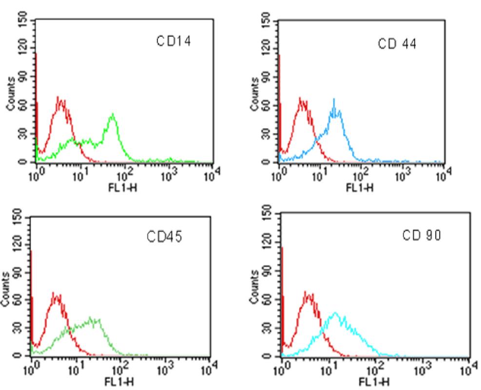
The amnion is a particular tissue whose cells show features of multipotent stem cells proposed for use in cellular therapy and regenerative medicine. From equine amnion collected after the foal birth we have isolated MSCs (mesenchymal stem cells), namely EAMSCs (equine amnion mesenchymal stem cells), from the mesoblastic layer. The cells were grown in α-MEM (α-modified minimum essential medium) and the effect of EGF (epidermal growth factor) supplementation was evaluated. To assess the growth kinetic of EAMSCs we have taken into account some parameters [PD (population doubling), fold increase and DT (doubling time)]. The differentiation in chondrogenic, adipogenic and osteogenic types of cells and their epitope expression by a cytofluorimetric study have been reported. EGF supplementation of the culture medium resulted in a significant increase in PD growth parameter and in the formation of bone nodules for the osteogenic differentiation. By immunohistochemistry the amnion tissue shows a positivity for the c-Kit (cluster tyrosine-protein kinase), CD105 and Oct-4 (octamer-binding transcription factor 4) antigens that confirmed the presence of MSCs with embryonic phenotype.


 分享
分享

 求助内容:
求助内容: 应助结果提醒方式:
 扫码关注我们
