{"title":"感觉诱发的皮层血流动力学反应的试验间变异性:刺激前波动幅度的作用。","authors":"Mohamad Saka, Jason Berwick, Myles Jones","doi":"10.3389/fnene.2012.00010","DOIUrl":null,"url":null,"abstract":"<p><p>Brain imaging techniques utilize hemodynamic changes that accompany brain activation. However, stimulus-evoked hemodynamic responses display considerable inter-trial variability and the sources of this variability are poorly understood. One of the sources of this response variation could be ongoing spontaneous hemodynamic fluctuations. We recently investigated this issue by measuring cortical hemodynamics in response to sensory stimuli in anesthetized rodents using 2-dimensional optical imaging spectroscopy. We suggested that sensory-evoked cortical hemodynamics displayed distinctive response characteristics and magnitudes depending on the phase of ongoing fluctuations at stimulus onset due to a linear superposition of evoked and ongoing hemodynamics (Saka et al., 2010). However, the previous analysis neglected to examine the possible influence of variability of the size of ongoing fluctuations. Consequently, data were further analyzed to examine whether the size of pre-stimulus hemodynamic fluctuations also influenced the magnitude of subsequent stimulus-evoked responses. Indeed, in the case of all individual trials, a moderate correlation between the size of the pre-stimulus fluctuations and the magnitudes of the subsequent sensory-evoked responses were observed. However, different correlations between the size of the pre-stimulus fluctuations and magnitudes of the subsequent sensory-evoked cortical hemodynamic responses could be observed depending on their phase at stimulus onset. These analyses suggest that both the size and phase of pre-stimulus fluctuations in cortical hemodynamics contribute to inter-trial variability in sensory-evoked responses.</p>","PeriodicalId":88242,"journal":{"name":"Frontiers in neuroenergetics","volume":"4 ","pages":"10"},"PeriodicalIF":0.0000,"publicationDate":"2012-11-05","publicationTypes":"Journal Article","fieldsOfStudy":null,"isOpenAccess":false,"openAccessPdf":"https://www.ncbi.nlm.nih.gov/pmc/articles/PMC3488699/pdf/","citationCount":"0","resultStr":"{\"title\":\"Inter-trial variability in sensory-evoked cortical hemodynamic responses: the role of the magnitude of pre-stimulus fluctuations.\",\"authors\":\"Mohamad Saka, Jason Berwick, Myles Jones\",\"doi\":\"10.3389/fnene.2012.00010\",\"DOIUrl\":null,\"url\":null,\"abstract\":\"<p><p>Brain imaging techniques utilize hemodynamic changes that accompany brain activation. However, stimulus-evoked hemodynamic responses display considerable inter-trial variability and the sources of this variability are poorly understood. One of the sources of this response variation could be ongoing spontaneous hemodynamic fluctuations. We recently investigated this issue by measuring cortical hemodynamics in response to sensory stimuli in anesthetized rodents using 2-dimensional optical imaging spectroscopy. We suggested that sensory-evoked cortical hemodynamics displayed distinctive response characteristics and magnitudes depending on the phase of ongoing fluctuations at stimulus onset due to a linear superposition of evoked and ongoing hemodynamics (Saka et al., 2010). However, the previous analysis neglected to examine the possible influence of variability of the size of ongoing fluctuations. Consequently, data were further analyzed to examine whether the size of pre-stimulus hemodynamic fluctuations also influenced the magnitude of subsequent stimulus-evoked responses. Indeed, in the case of all individual trials, a moderate correlation between the size of the pre-stimulus fluctuations and the magnitudes of the subsequent sensory-evoked responses were observed. However, different correlations between the size of the pre-stimulus fluctuations and magnitudes of the subsequent sensory-evoked cortical hemodynamic responses could be observed depending on their phase at stimulus onset. These analyses suggest that both the size and phase of pre-stimulus fluctuations in cortical hemodynamics contribute to inter-trial variability in sensory-evoked responses.</p>\",\"PeriodicalId\":88242,\"journal\":{\"name\":\"Frontiers in neuroenergetics\",\"volume\":\"4 \",\"pages\":\"10\"},\"PeriodicalIF\":0.0000,\"publicationDate\":\"2012-11-05\",\"publicationTypes\":\"Journal Article\",\"fieldsOfStudy\":null,\"isOpenAccess\":false,\"openAccessPdf\":\"https://www.ncbi.nlm.nih.gov/pmc/articles/PMC3488699/pdf/\",\"citationCount\":\"0\",\"resultStr\":null,\"platform\":\"Semanticscholar\",\"paperid\":null,\"PeriodicalName\":\"Frontiers in neuroenergetics\",\"FirstCategoryId\":\"1085\",\"ListUrlMain\":\"https://doi.org/10.3389/fnene.2012.00010\",\"RegionNum\":0,\"RegionCategory\":null,\"ArticlePicture\":[],\"TitleCN\":null,\"AbstractTextCN\":null,\"PMCID\":null,\"EPubDate\":\"2012/1/1 0:00:00\",\"PubModel\":\"eCollection\",\"JCR\":\"\",\"JCRName\":\"\",\"Score\":null,\"Total\":0}","platform":"Semanticscholar","paperid":null,"PeriodicalName":"Frontiers in neuroenergetics","FirstCategoryId":"1085","ListUrlMain":"https://doi.org/10.3389/fnene.2012.00010","RegionNum":0,"RegionCategory":null,"ArticlePicture":[],"TitleCN":null,"AbstractTextCN":null,"PMCID":null,"EPubDate":"2012/1/1 0:00:00","PubModel":"eCollection","JCR":"","JCRName":"","Score":null,"Total":0}
Inter-trial variability in sensory-evoked cortical hemodynamic responses: the role of the magnitude of pre-stimulus fluctuations.
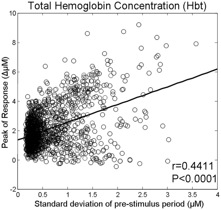
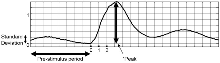
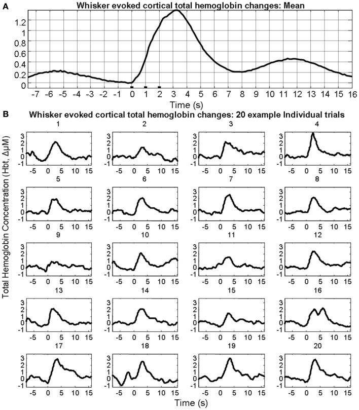
Brain imaging techniques utilize hemodynamic changes that accompany brain activation. However, stimulus-evoked hemodynamic responses display considerable inter-trial variability and the sources of this variability are poorly understood. One of the sources of this response variation could be ongoing spontaneous hemodynamic fluctuations. We recently investigated this issue by measuring cortical hemodynamics in response to sensory stimuli in anesthetized rodents using 2-dimensional optical imaging spectroscopy. We suggested that sensory-evoked cortical hemodynamics displayed distinctive response characteristics and magnitudes depending on the phase of ongoing fluctuations at stimulus onset due to a linear superposition of evoked and ongoing hemodynamics (Saka et al., 2010). However, the previous analysis neglected to examine the possible influence of variability of the size of ongoing fluctuations. Consequently, data were further analyzed to examine whether the size of pre-stimulus hemodynamic fluctuations also influenced the magnitude of subsequent stimulus-evoked responses. Indeed, in the case of all individual trials, a moderate correlation between the size of the pre-stimulus fluctuations and the magnitudes of the subsequent sensory-evoked responses were observed. However, different correlations between the size of the pre-stimulus fluctuations and magnitudes of the subsequent sensory-evoked cortical hemodynamic responses could be observed depending on their phase at stimulus onset. These analyses suggest that both the size and phase of pre-stimulus fluctuations in cortical hemodynamics contribute to inter-trial variability in sensory-evoked responses.


 分享
分享

 求助内容:
求助内容: 应助结果提醒方式:
 扫码关注我们
