Mostafa A Borahay, Fangxian Lu, Bulent Ozpolat, Ibrahim Tekedereli, Bilgin Gurates, Sinem Karipcin, Gokhan S Kilic
{"title":"苗勒氏管抑制物质可抑制子宫内膜异位症体外细胞的增殖并诱导其凋亡和自噬。","authors":"Mostafa A Borahay, Fangxian Lu, Bulent Ozpolat, Ibrahim Tekedereli, Bilgin Gurates, Sinem Karipcin, Gokhan S Kilic","doi":"10.1155/2013/361489","DOIUrl":null,"url":null,"abstract":"<p><p>Objective. To determine the effects of Mullerian inhibiting substance (MIS) treatment on endometriosis cells through study of apoptosis and autophagy. Design. Experimental in vitro study. Setting. University research laboratory. Cell Line. CRL-7566 endometriosis cell line. This line was established from a benign ovarian cyst taken from a patient with endometriosis. Interventions. In vitro treatment with MIS. Main Outcome Measures. The main outcome measures were cellular viability, proliferation, cell-cycle arrest, and induction of apoptosis and autophagy in endometriotic cells. Results. MIS treatment inhibited proliferation of endometriosis cells and induced apoptosis, as indicated by Annexin V staining, and induced caspase-9 cleavage and cell-cycle arrest, as evidenced by increased expression of p27 CDK-inhibitor. MIS treatment also induced autophagy in endometriosis cells as demonstrated by a significant increase in LC3-II induction, a hallmark of autophagy. Conclusions. MIS inhibits cell growth and induces autophagy, as well as apoptosis, in ectopic endometrial cell lines. Our results suggest that MIS may have a potential as a novel approach for medical treatment of endometriosis. Further studies may be needed to test the efficacy of MIS treatment in animal models and to develop MIS treatment specifically targeted to the endometriosis. </p>","PeriodicalId":73520,"journal":{"name":"ISRN obstetrics and gynecology","volume":"2013 ","pages":"361489"},"PeriodicalIF":0.0000,"publicationDate":"2013-06-19","publicationTypes":"Journal Article","fieldsOfStudy":null,"isOpenAccess":false,"openAccessPdf":"https://www.ncbi.nlm.nih.gov/pmc/articles/PMC3703732/pdf/","citationCount":"0","resultStr":"{\"title\":\"Mullerian inhibiting substance suppresses proliferation and induces apoptosis and autophagy in endometriosis cells in vitro.\",\"authors\":\"Mostafa A Borahay, Fangxian Lu, Bulent Ozpolat, Ibrahim Tekedereli, Bilgin Gurates, Sinem Karipcin, Gokhan S Kilic\",\"doi\":\"10.1155/2013/361489\",\"DOIUrl\":null,\"url\":null,\"abstract\":\"<p><p>Objective. To determine the effects of Mullerian inhibiting substance (MIS) treatment on endometriosis cells through study of apoptosis and autophagy. Design. Experimental in vitro study. Setting. University research laboratory. Cell Line. CRL-7566 endometriosis cell line. This line was established from a benign ovarian cyst taken from a patient with endometriosis. Interventions. In vitro treatment with MIS. Main Outcome Measures. The main outcome measures were cellular viability, proliferation, cell-cycle arrest, and induction of apoptosis and autophagy in endometriotic cells. Results. MIS treatment inhibited proliferation of endometriosis cells and induced apoptosis, as indicated by Annexin V staining, and induced caspase-9 cleavage and cell-cycle arrest, as evidenced by increased expression of p27 CDK-inhibitor. MIS treatment also induced autophagy in endometriosis cells as demonstrated by a significant increase in LC3-II induction, a hallmark of autophagy. Conclusions. MIS inhibits cell growth and induces autophagy, as well as apoptosis, in ectopic endometrial cell lines. Our results suggest that MIS may have a potential as a novel approach for medical treatment of endometriosis. Further studies may be needed to test the efficacy of MIS treatment in animal models and to develop MIS treatment specifically targeted to the endometriosis. </p>\",\"PeriodicalId\":73520,\"journal\":{\"name\":\"ISRN obstetrics and gynecology\",\"volume\":\"2013 \",\"pages\":\"361489\"},\"PeriodicalIF\":0.0000,\"publicationDate\":\"2013-06-19\",\"publicationTypes\":\"Journal Article\",\"fieldsOfStudy\":null,\"isOpenAccess\":false,\"openAccessPdf\":\"https://www.ncbi.nlm.nih.gov/pmc/articles/PMC3703732/pdf/\",\"citationCount\":\"0\",\"resultStr\":null,\"platform\":\"Semanticscholar\",\"paperid\":null,\"PeriodicalName\":\"ISRN obstetrics and gynecology\",\"FirstCategoryId\":\"1085\",\"ListUrlMain\":\"https://doi.org/10.1155/2013/361489\",\"RegionNum\":0,\"RegionCategory\":null,\"ArticlePicture\":[],\"TitleCN\":null,\"AbstractTextCN\":null,\"PMCID\":null,\"EPubDate\":\"2013/1/1 0:00:00\",\"PubModel\":\"Print\",\"JCR\":\"\",\"JCRName\":\"\",\"Score\":null,\"Total\":0}","platform":"Semanticscholar","paperid":null,"PeriodicalName":"ISRN obstetrics and gynecology","FirstCategoryId":"1085","ListUrlMain":"https://doi.org/10.1155/2013/361489","RegionNum":0,"RegionCategory":null,"ArticlePicture":[],"TitleCN":null,"AbstractTextCN":null,"PMCID":null,"EPubDate":"2013/1/1 0:00:00","PubModel":"Print","JCR":"","JCRName":"","Score":null,"Total":0}
引用次数: 0
摘要
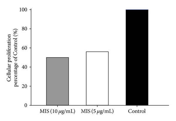
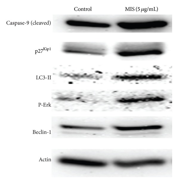
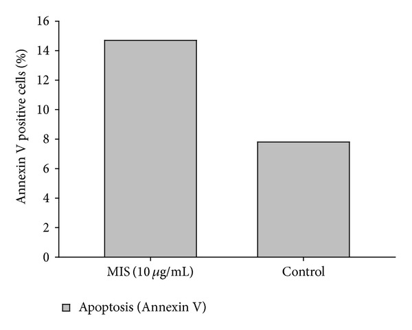
目的通过研究细胞凋亡和自噬,确定穆勒氏管抑制物质(MIS)治疗对子宫内膜异位症细胞的影响。设计。体外实验研究。环境。大学研究实验室。细胞系。CRL-7566 子宫内膜异位症细胞系。该细胞系取自一名子宫内膜异位症患者的良性卵巢囊肿。干预措施。用 MIS 进行体外治疗。主要结果指标。主要结果指标为子宫内膜异位细胞的细胞活力、增殖、细胞周期停滞、诱导凋亡和自噬。结果MIS 治疗可抑制子宫内膜异位症细胞的增殖,诱导细胞凋亡(Annexin V 染色显示),诱导 caspase-9 分裂和细胞周期停滞(p27 CDK 抑制剂表达增加显示)。MIS 处理还能诱导子宫内膜异位症细胞自噬,自噬的标志 LC3-II 的诱导量显著增加就是证明。结论MIS 可抑制异位子宫内膜细胞系的细胞生长,诱导自噬和细胞凋亡。我们的研究结果表明,MIS 有可能成为治疗子宫内膜异位症的一种新方法。可能还需要进一步的研究来检验 MIS 在动物模型中的疗效,并开发出专门针对子宫内膜异位症的 MIS 治疗方法。
Mullerian inhibiting substance suppresses proliferation and induces apoptosis and autophagy in endometriosis cells in vitro.
Objective. To determine the effects of Mullerian inhibiting substance (MIS) treatment on endometriosis cells through study of apoptosis and autophagy. Design. Experimental in vitro study. Setting. University research laboratory. Cell Line. CRL-7566 endometriosis cell line. This line was established from a benign ovarian cyst taken from a patient with endometriosis. Interventions. In vitro treatment with MIS. Main Outcome Measures. The main outcome measures were cellular viability, proliferation, cell-cycle arrest, and induction of apoptosis and autophagy in endometriotic cells. Results. MIS treatment inhibited proliferation of endometriosis cells and induced apoptosis, as indicated by Annexin V staining, and induced caspase-9 cleavage and cell-cycle arrest, as evidenced by increased expression of p27 CDK-inhibitor. MIS treatment also induced autophagy in endometriosis cells as demonstrated by a significant increase in LC3-II induction, a hallmark of autophagy. Conclusions. MIS inhibits cell growth and induces autophagy, as well as apoptosis, in ectopic endometrial cell lines. Our results suggest that MIS may have a potential as a novel approach for medical treatment of endometriosis. Further studies may be needed to test the efficacy of MIS treatment in animal models and to develop MIS treatment specifically targeted to the endometriosis.


 分享
分享

 求助内容:
求助内容: 应助结果提醒方式:
 扫码关注我们
