{"title":"用力谱法研究了TiO2细颗粒和纳米粒子聚集体对人体中性粒细胞纳米力学性能的动力学影响。","authors":"Everton Luis Santos da Rosa","doi":"10.1186/2046-1682-6-11","DOIUrl":null,"url":null,"abstract":"<p><strong>Background: </strong>Increasing applications of titanium dioxide (TiO2) fine particles (FPs) and nanoparticles (NPs) require coupled knowledge improvement concerning their biokinetic effects. Neutrophils are quickly recruited to titanium implantation areas. Neutrophils mechanical properties display a crucial role on cell physiology and immune responsive functions. Then, micro and nanomechanical characterization assessed by force spectroscopy (FS) technique has been largely applied in this field.</p><p><strong>Results: </strong>Scanning electron microscopy (SEM) images highlighted neutrophils morphological changes along TiO2 FPs and NPs aggregates exposure time (1, 5, and 30 min) compared to controls. FS approaches showed an increasing on attraction forces to TiO2 FPs and NPs treated neutrophils. This group depicted stronger stiffness features than controls just at 1 min of exposure. Treated neutrophils showed a tendency to increase adhesive properties after 1 and 5 min of exposure. These cells maintained comparatively higher elasticity behavior for a longer time possibly due to intense phagocytosis and cell stiffness opposing to the tip indentation. Neutrophils activation caused by FPs and NPs uptake could be related to increasing dissipated energy results.</p><p><strong>Conclusions: </strong>Mechanical modifications resulted from TiO2 FPs and NPs aggregates interaction with neutrophils showed increasing stiffness and also cell morphology alteration. Cells treatment by this metal FPs and NPs caused an increase in attractive forces. This event was mainly observed on the initial exposure times probably regarding to the interaction of neutrophils membrane and phagocytosis. Similar results were found to adhesion forces and dissipated energy outcomes. Treated cells presented comparatively higher elasticity behavior for a longer time. SEM images clearly suggested cell morphology alteration along time course probably related to activation, cytoskeleton rearrangement and phagocytosis. This scenario with increase in stiffness strongly suggests a direct relationship over neutrophil rolling, arrest, and transmigration. Scrutinizing these interactions represents an essential step to clarify the mechanisms involved on treatments containing micro and nanomaterials and their fates on the organisms.</p>","PeriodicalId":9045,"journal":{"name":"BMC Biophysics","volume":"6 1","pages":"11"},"PeriodicalIF":0.0000,"publicationDate":"2013-08-19","publicationTypes":"Journal Article","fieldsOfStudy":null,"isOpenAccess":false,"openAccessPdf":"https://sci-hub-pdf.com/10.1186/2046-1682-6-11","citationCount":"15","resultStr":"{\"title\":\"Kinetic effects of TiO2 fine particles and nanoparticles aggregates on the nanomechanical properties of human neutrophils assessed by force spectroscopy.\",\"authors\":\"Everton Luis Santos da Rosa\",\"doi\":\"10.1186/2046-1682-6-11\",\"DOIUrl\":null,\"url\":null,\"abstract\":\"<p><strong>Background: </strong>Increasing applications of titanium dioxide (TiO2) fine particles (FPs) and nanoparticles (NPs) require coupled knowledge improvement concerning their biokinetic effects. Neutrophils are quickly recruited to titanium implantation areas. Neutrophils mechanical properties display a crucial role on cell physiology and immune responsive functions. Then, micro and nanomechanical characterization assessed by force spectroscopy (FS) technique has been largely applied in this field.</p><p><strong>Results: </strong>Scanning electron microscopy (SEM) images highlighted neutrophils morphological changes along TiO2 FPs and NPs aggregates exposure time (1, 5, and 30 min) compared to controls. FS approaches showed an increasing on attraction forces to TiO2 FPs and NPs treated neutrophils. This group depicted stronger stiffness features than controls just at 1 min of exposure. Treated neutrophils showed a tendency to increase adhesive properties after 1 and 5 min of exposure. These cells maintained comparatively higher elasticity behavior for a longer time possibly due to intense phagocytosis and cell stiffness opposing to the tip indentation. Neutrophils activation caused by FPs and NPs uptake could be related to increasing dissipated energy results.</p><p><strong>Conclusions: </strong>Mechanical modifications resulted from TiO2 FPs and NPs aggregates interaction with neutrophils showed increasing stiffness and also cell morphology alteration. Cells treatment by this metal FPs and NPs caused an increase in attractive forces. This event was mainly observed on the initial exposure times probably regarding to the interaction of neutrophils membrane and phagocytosis. Similar results were found to adhesion forces and dissipated energy outcomes. Treated cells presented comparatively higher elasticity behavior for a longer time. SEM images clearly suggested cell morphology alteration along time course probably related to activation, cytoskeleton rearrangement and phagocytosis. This scenario with increase in stiffness strongly suggests a direct relationship over neutrophil rolling, arrest, and transmigration. Scrutinizing these interactions represents an essential step to clarify the mechanisms involved on treatments containing micro and nanomaterials and their fates on the organisms.</p>\",\"PeriodicalId\":9045,\"journal\":{\"name\":\"BMC Biophysics\",\"volume\":\"6 1\",\"pages\":\"11\"},\"PeriodicalIF\":0.0000,\"publicationDate\":\"2013-08-19\",\"publicationTypes\":\"Journal Article\",\"fieldsOfStudy\":null,\"isOpenAccess\":false,\"openAccessPdf\":\"https://sci-hub-pdf.com/10.1186/2046-1682-6-11\",\"citationCount\":\"15\",\"resultStr\":null,\"platform\":\"Semanticscholar\",\"paperid\":null,\"PeriodicalName\":\"BMC Biophysics\",\"FirstCategoryId\":\"1085\",\"ListUrlMain\":\"https://doi.org/10.1186/2046-1682-6-11\",\"RegionNum\":0,\"RegionCategory\":null,\"ArticlePicture\":[],\"TitleCN\":null,\"AbstractTextCN\":null,\"PMCID\":null,\"EPubDate\":\"\",\"PubModel\":\"\",\"JCR\":\"Q1\",\"JCRName\":\"Biochemistry, Genetics and Molecular Biology\",\"Score\":null,\"Total\":0}","platform":"Semanticscholar","paperid":null,"PeriodicalName":"BMC Biophysics","FirstCategoryId":"1085","ListUrlMain":"https://doi.org/10.1186/2046-1682-6-11","RegionNum":0,"RegionCategory":null,"ArticlePicture":[],"TitleCN":null,"AbstractTextCN":null,"PMCID":null,"EPubDate":"","PubModel":"","JCR":"Q1","JCRName":"Biochemistry, Genetics and Molecular Biology","Score":null,"Total":0}
Kinetic effects of TiO2 fine particles and nanoparticles aggregates on the nanomechanical properties of human neutrophils assessed by force spectroscopy.
Background: Increasing applications of titanium dioxide (TiO2) fine particles (FPs) and nanoparticles (NPs) require coupled knowledge improvement concerning their biokinetic effects. Neutrophils are quickly recruited to titanium implantation areas. Neutrophils mechanical properties display a crucial role on cell physiology and immune responsive functions. Then, micro and nanomechanical characterization assessed by force spectroscopy (FS) technique has been largely applied in this field.
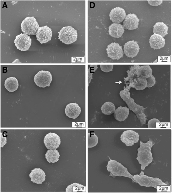
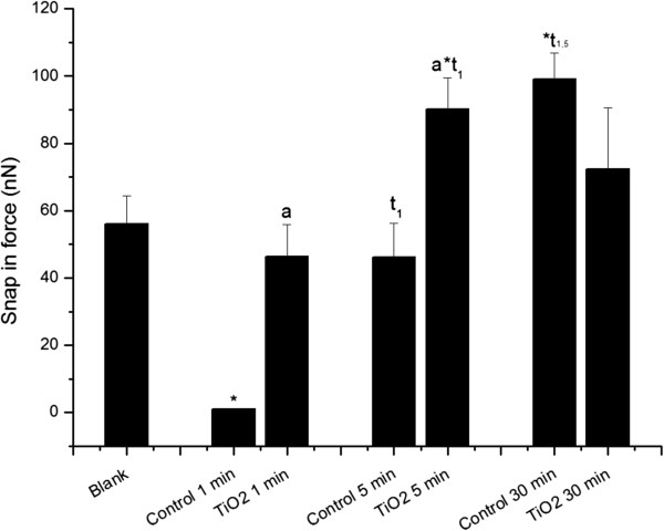
Results: Scanning electron microscopy (SEM) images highlighted neutrophils morphological changes along TiO2 FPs and NPs aggregates exposure time (1, 5, and 30 min) compared to controls. FS approaches showed an increasing on attraction forces to TiO2 FPs and NPs treated neutrophils. This group depicted stronger stiffness features than controls just at 1 min of exposure. Treated neutrophils showed a tendency to increase adhesive properties after 1 and 5 min of exposure. These cells maintained comparatively higher elasticity behavior for a longer time possibly due to intense phagocytosis and cell stiffness opposing to the tip indentation. Neutrophils activation caused by FPs and NPs uptake could be related to increasing dissipated energy results.
Conclusions: Mechanical modifications resulted from TiO2 FPs and NPs aggregates interaction with neutrophils showed increasing stiffness and also cell morphology alteration. Cells treatment by this metal FPs and NPs caused an increase in attractive forces. This event was mainly observed on the initial exposure times probably regarding to the interaction of neutrophils membrane and phagocytosis. Similar results were found to adhesion forces and dissipated energy outcomes. Treated cells presented comparatively higher elasticity behavior for a longer time. SEM images clearly suggested cell morphology alteration along time course probably related to activation, cytoskeleton rearrangement and phagocytosis. This scenario with increase in stiffness strongly suggests a direct relationship over neutrophil rolling, arrest, and transmigration. Scrutinizing these interactions represents an essential step to clarify the mechanisms involved on treatments containing micro and nanomaterials and their fates on the organisms.


 分享
分享
 求助内容:
求助内容: 应助结果提醒方式:
 扫码关注我们
