{"title":"磁共振成像(MRI)在帕金森病中的应用。","authors":"Paul J Tuite, Silvia Mangia, Shalom Michaeli","doi":"10.4172/2161-0460.S1-001","DOIUrl":null,"url":null,"abstract":"<p><p>Recent developments in brain imaging methods are on the verge of changing the evaluation of people with Parkinson's disease (PD). This includes an assortment of techniques ranging from diffusion tensor imaging (DTI) to iron-sensitive methods such as T<sub>2</sub><sup>*</sup>, as well as adiabatic methods R<sub>1ρ</sub> and R<sub>2ρ</sub>, resting-state functional MRI, and magnetic resonance spectroscopy (MRS). Using a multi-modality approach that ascertains different aspects of the pathophysiology or pathology of PD, it may be possible to better characterize disease phenotypes as well as provide a surrogate of disease and a potential means to track disease progression.</p>","PeriodicalId":15013,"journal":{"name":"Journal of Alzheimer's disease & Parkinsonism","volume":"Suppl 1 ","pages":"001"},"PeriodicalIF":0.0000,"publicationDate":"2013-03-25","publicationTypes":"Journal Article","fieldsOfStudy":null,"isOpenAccess":false,"openAccessPdf":"https://sci-hub-pdf.com/10.4172/2161-0460.S1-001","citationCount":"32","resultStr":"{\"title\":\"Magnetic Resonance Imaging (MRI) in Parkinson's Disease.\",\"authors\":\"Paul J Tuite, Silvia Mangia, Shalom Michaeli\",\"doi\":\"10.4172/2161-0460.S1-001\",\"DOIUrl\":null,\"url\":null,\"abstract\":\"<p><p>Recent developments in brain imaging methods are on the verge of changing the evaluation of people with Parkinson's disease (PD). This includes an assortment of techniques ranging from diffusion tensor imaging (DTI) to iron-sensitive methods such as T<sub>2</sub><sup>*</sup>, as well as adiabatic methods R<sub>1ρ</sub> and R<sub>2ρ</sub>, resting-state functional MRI, and magnetic resonance spectroscopy (MRS). Using a multi-modality approach that ascertains different aspects of the pathophysiology or pathology of PD, it may be possible to better characterize disease phenotypes as well as provide a surrogate of disease and a potential means to track disease progression.</p>\",\"PeriodicalId\":15013,\"journal\":{\"name\":\"Journal of Alzheimer's disease & Parkinsonism\",\"volume\":\"Suppl 1 \",\"pages\":\"001\"},\"PeriodicalIF\":0.0000,\"publicationDate\":\"2013-03-25\",\"publicationTypes\":\"Journal Article\",\"fieldsOfStudy\":null,\"isOpenAccess\":false,\"openAccessPdf\":\"https://sci-hub-pdf.com/10.4172/2161-0460.S1-001\",\"citationCount\":\"32\",\"resultStr\":null,\"platform\":\"Semanticscholar\",\"paperid\":null,\"PeriodicalName\":\"Journal of Alzheimer's disease & Parkinsonism\",\"FirstCategoryId\":\"1085\",\"ListUrlMain\":\"https://doi.org/10.4172/2161-0460.S1-001\",\"RegionNum\":0,\"RegionCategory\":null,\"ArticlePicture\":[],\"TitleCN\":null,\"AbstractTextCN\":null,\"PMCID\":null,\"EPubDate\":\"\",\"PubModel\":\"\",\"JCR\":\"\",\"JCRName\":\"\",\"Score\":null,\"Total\":0}","platform":"Semanticscholar","paperid":null,"PeriodicalName":"Journal of Alzheimer's disease & Parkinsonism","FirstCategoryId":"1085","ListUrlMain":"https://doi.org/10.4172/2161-0460.S1-001","RegionNum":0,"RegionCategory":null,"ArticlePicture":[],"TitleCN":null,"AbstractTextCN":null,"PMCID":null,"EPubDate":"","PubModel":"","JCR":"","JCRName":"","Score":null,"Total":0}
Magnetic Resonance Imaging (MRI) in Parkinson's Disease.
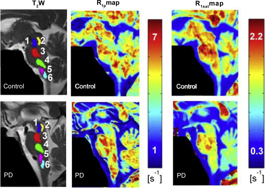
Recent developments in brain imaging methods are on the verge of changing the evaluation of people with Parkinson's disease (PD). This includes an assortment of techniques ranging from diffusion tensor imaging (DTI) to iron-sensitive methods such as T2*, as well as adiabatic methods R1ρ and R2ρ, resting-state functional MRI, and magnetic resonance spectroscopy (MRS). Using a multi-modality approach that ascertains different aspects of the pathophysiology or pathology of PD, it may be possible to better characterize disease phenotypes as well as provide a surrogate of disease and a potential means to track disease progression.


 分享
分享

 求助内容:
求助内容: 应助结果提醒方式:
 扫码关注我们
