{"title":"抑制剂配合物中S6K1激酶结构域的晶体结构。","authors":"Hideaki Niwa, Junko Mikuni, Shunta Sasaki, Yuri Tomabechi, Keiko Honda, Mariko Ikeda, Noboru Ohsawa, Motoaki Wakiyama, Noriko Handa, Mikako Shirouzu, Teruki Honma, Akiko Tanaka, Shigeyuki Yokoyama","doi":"10.1007/s10969-014-9188-8","DOIUrl":null,"url":null,"abstract":"<p><p>Ribosomal protein S6 kinase 1 (S6K1) is a serine/threonine protein kinase that plays an important role in the PIK3/mTOR signaling pathway, and is implicated in diseases including diabetes, obesity, and cancer. The crystal structures of the S6K1 kinase domain in complexes with staurosporine and the S6K1-specific inhibitor PF-4708671 have been reported. In the present study, five compounds (F108, F109, F176, F177, and F179) were newly identified by in silico screening of a chemical library and kinase assay. The crystal structures of the five inhibitors in complexes with the S6K1 kinase domain were determined at resolutions between 1.85 and 2.10 Å. All of the inhibitors bound to the ATP binding site, lying along the P-loop, while the activation loop stayed in the inactive form. Compound F179, with a carbonyl group in the middle of the molecule, altered the αC helix conformation by interacting with the invariant Lys123. Compounds F176 and F177 bound slightly distant from the hinge region, and their sulfoamide groups formed polar interactions with the protein. The structural features required for the specific binding of inhibitors are discussed.</p>","PeriodicalId":73957,"journal":{"name":"Journal of structural and functional genomics","volume":" ","pages":"153-64"},"PeriodicalIF":0.0000,"publicationDate":"2014-09-01","publicationTypes":"Journal Article","fieldsOfStudy":null,"isOpenAccess":false,"openAccessPdf":"https://sci-hub-pdf.com/10.1007/s10969-014-9188-8","citationCount":"13","resultStr":"{\"title\":\"Crystal structures of the S6K1 kinase domain in complexes with inhibitors.\",\"authors\":\"Hideaki Niwa, Junko Mikuni, Shunta Sasaki, Yuri Tomabechi, Keiko Honda, Mariko Ikeda, Noboru Ohsawa, Motoaki Wakiyama, Noriko Handa, Mikako Shirouzu, Teruki Honma, Akiko Tanaka, Shigeyuki Yokoyama\",\"doi\":\"10.1007/s10969-014-9188-8\",\"DOIUrl\":null,\"url\":null,\"abstract\":\"<p><p>Ribosomal protein S6 kinase 1 (S6K1) is a serine/threonine protein kinase that plays an important role in the PIK3/mTOR signaling pathway, and is implicated in diseases including diabetes, obesity, and cancer. The crystal structures of the S6K1 kinase domain in complexes with staurosporine and the S6K1-specific inhibitor PF-4708671 have been reported. In the present study, five compounds (F108, F109, F176, F177, and F179) were newly identified by in silico screening of a chemical library and kinase assay. The crystal structures of the five inhibitors in complexes with the S6K1 kinase domain were determined at resolutions between 1.85 and 2.10 Å. All of the inhibitors bound to the ATP binding site, lying along the P-loop, while the activation loop stayed in the inactive form. Compound F179, with a carbonyl group in the middle of the molecule, altered the αC helix conformation by interacting with the invariant Lys123. Compounds F176 and F177 bound slightly distant from the hinge region, and their sulfoamide groups formed polar interactions with the protein. The structural features required for the specific binding of inhibitors are discussed.</p>\",\"PeriodicalId\":73957,\"journal\":{\"name\":\"Journal of structural and functional genomics\",\"volume\":\" \",\"pages\":\"153-64\"},\"PeriodicalIF\":0.0000,\"publicationDate\":\"2014-09-01\",\"publicationTypes\":\"Journal Article\",\"fieldsOfStudy\":null,\"isOpenAccess\":false,\"openAccessPdf\":\"https://sci-hub-pdf.com/10.1007/s10969-014-9188-8\",\"citationCount\":\"13\",\"resultStr\":null,\"platform\":\"Semanticscholar\",\"paperid\":null,\"PeriodicalName\":\"Journal of structural and functional genomics\",\"FirstCategoryId\":\"1085\",\"ListUrlMain\":\"https://doi.org/10.1007/s10969-014-9188-8\",\"RegionNum\":0,\"RegionCategory\":null,\"ArticlePicture\":[],\"TitleCN\":null,\"AbstractTextCN\":null,\"PMCID\":null,\"EPubDate\":\"2014/7/31 0:00:00\",\"PubModel\":\"Epub\",\"JCR\":\"\",\"JCRName\":\"\",\"Score\":null,\"Total\":0}","platform":"Semanticscholar","paperid":null,"PeriodicalName":"Journal of structural and functional genomics","FirstCategoryId":"1085","ListUrlMain":"https://doi.org/10.1007/s10969-014-9188-8","RegionNum":0,"RegionCategory":null,"ArticlePicture":[],"TitleCN":null,"AbstractTextCN":null,"PMCID":null,"EPubDate":"2014/7/31 0:00:00","PubModel":"Epub","JCR":"","JCRName":"","Score":null,"Total":0}
Crystal structures of the S6K1 kinase domain in complexes with inhibitors.
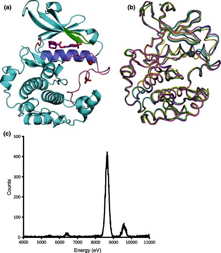
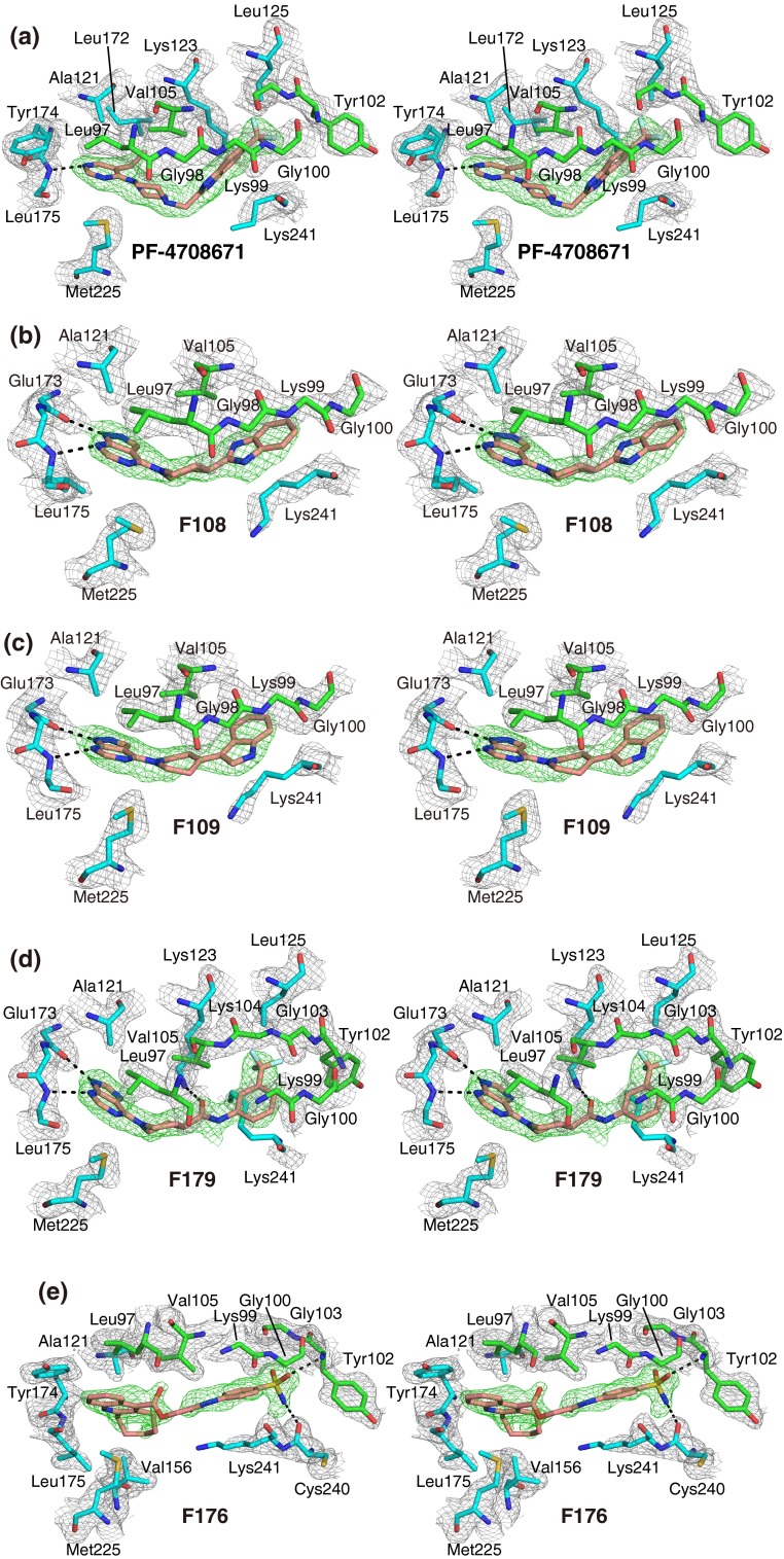
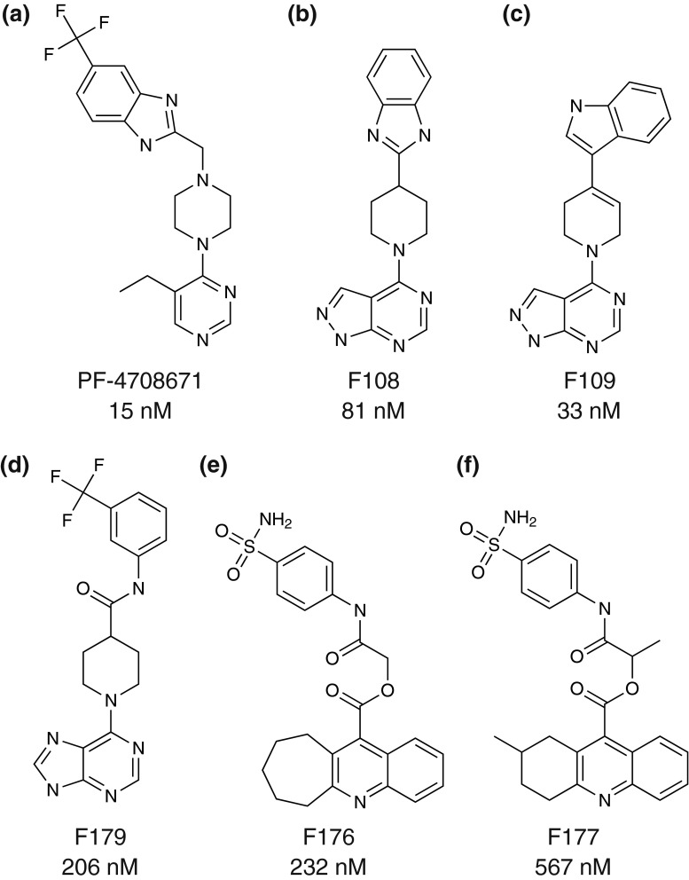
Ribosomal protein S6 kinase 1 (S6K1) is a serine/threonine protein kinase that plays an important role in the PIK3/mTOR signaling pathway, and is implicated in diseases including diabetes, obesity, and cancer. The crystal structures of the S6K1 kinase domain in complexes with staurosporine and the S6K1-specific inhibitor PF-4708671 have been reported. In the present study, five compounds (F108, F109, F176, F177, and F179) were newly identified by in silico screening of a chemical library and kinase assay. The crystal structures of the five inhibitors in complexes with the S6K1 kinase domain were determined at resolutions between 1.85 and 2.10 Å. All of the inhibitors bound to the ATP binding site, lying along the P-loop, while the activation loop stayed in the inactive form. Compound F179, with a carbonyl group in the middle of the molecule, altered the αC helix conformation by interacting with the invariant Lys123. Compounds F176 and F177 bound slightly distant from the hinge region, and their sulfoamide groups formed polar interactions with the protein. The structural features required for the specific binding of inhibitors are discussed.


 分享
分享

 求助内容:
求助内容: 应助结果提醒方式:
 扫码关注我们
